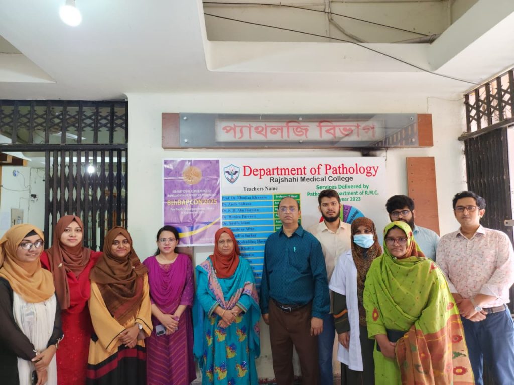
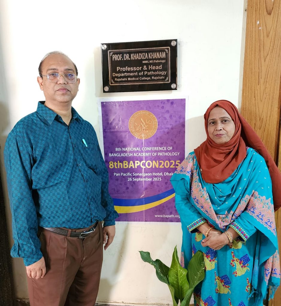
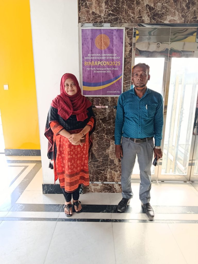
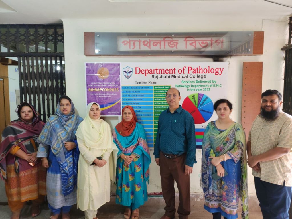
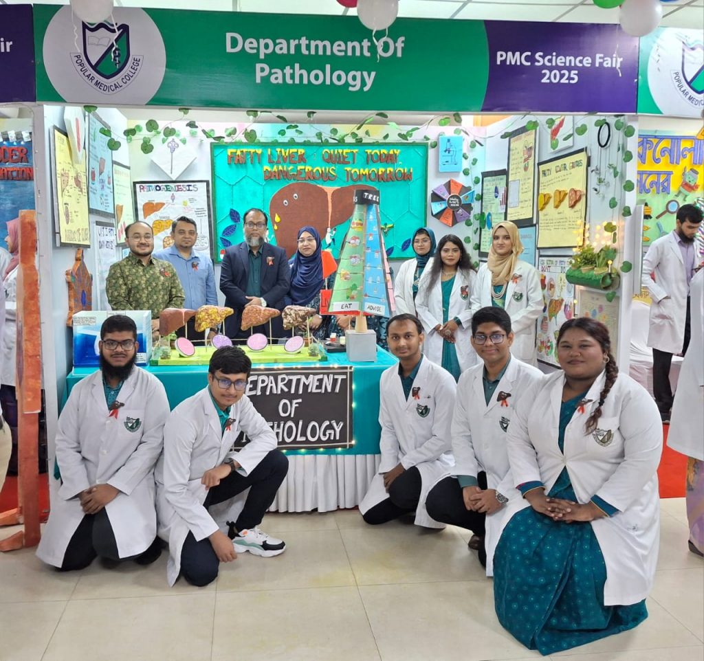
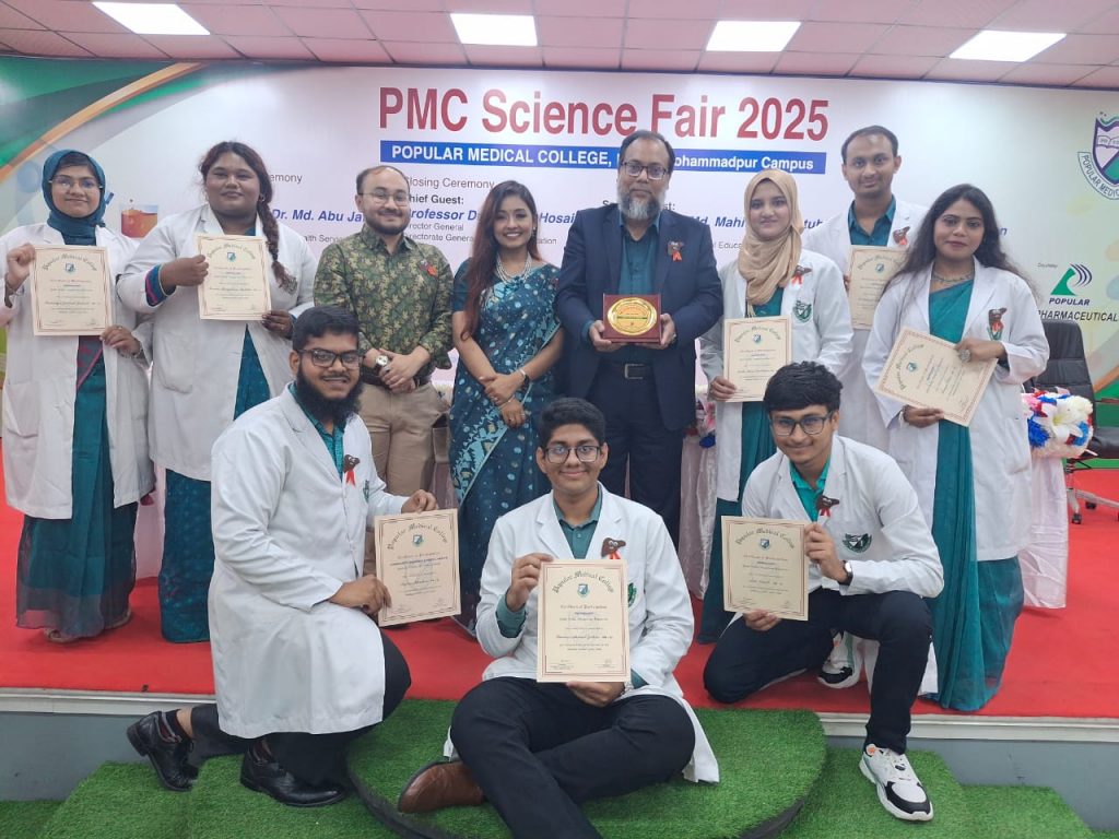
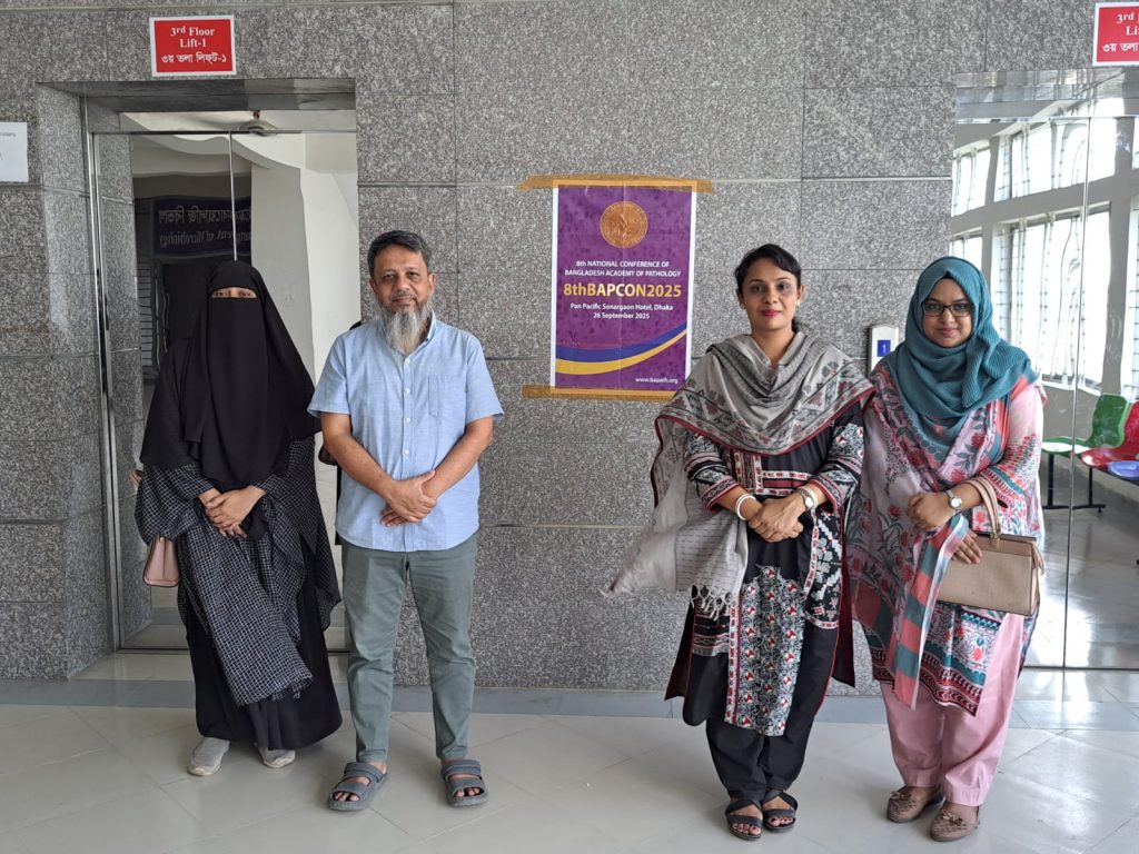
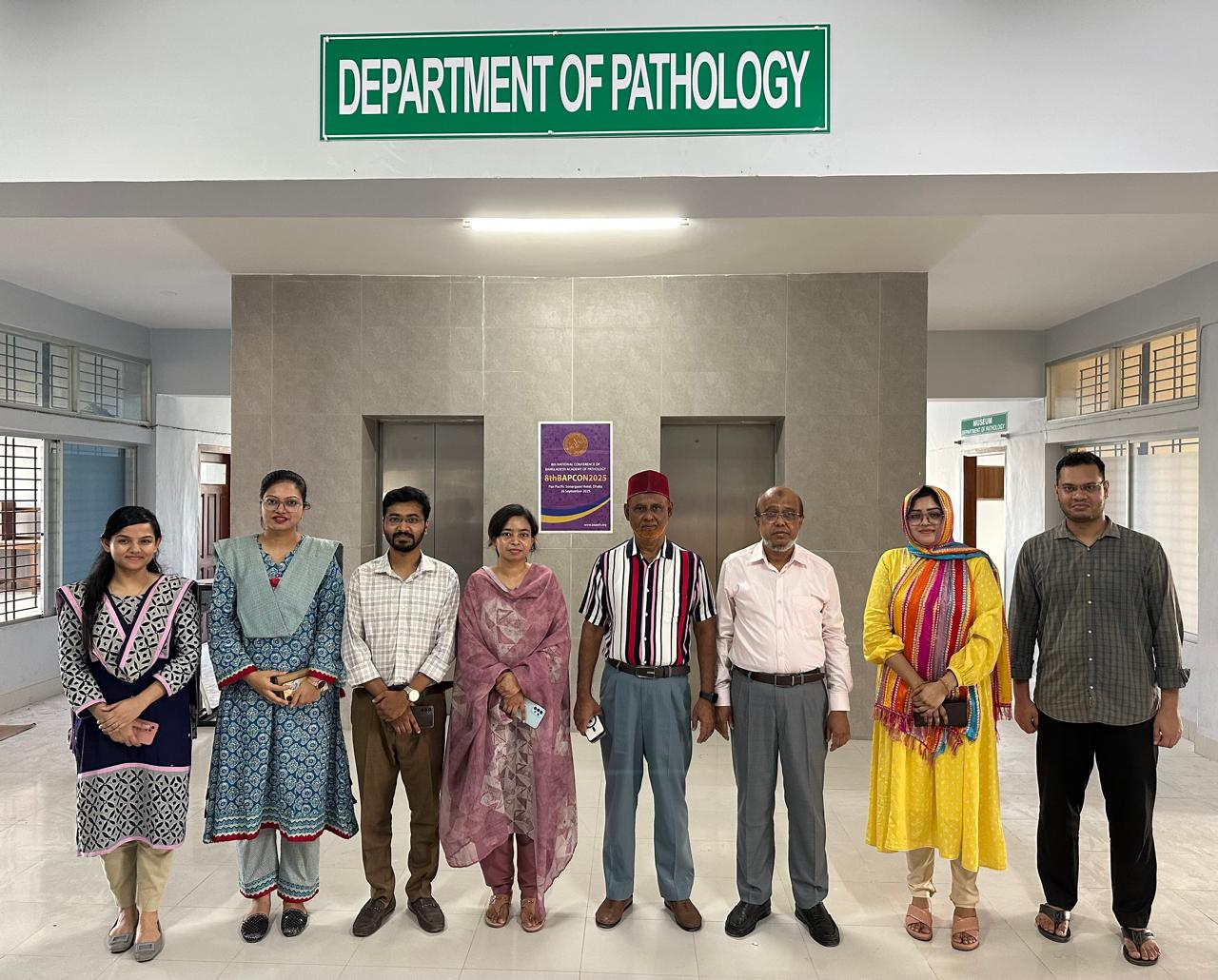
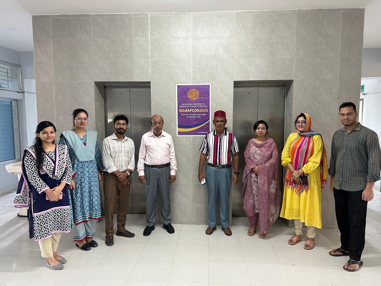
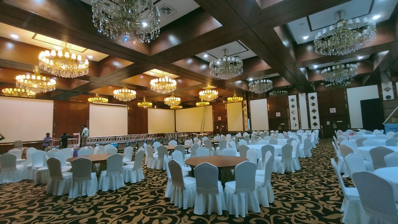
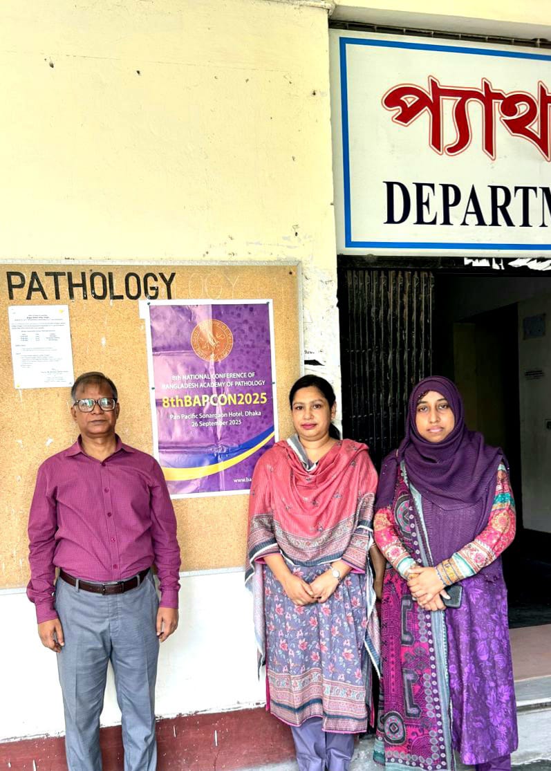
8thbapcon2025 Related Pictures


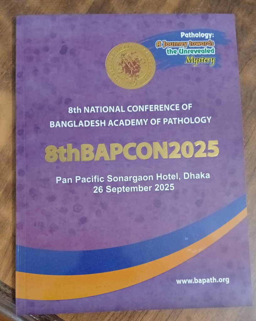


Bangladesh Academy of Pathology
Official Website of Bangladesh Academy of Pathology (BAP)
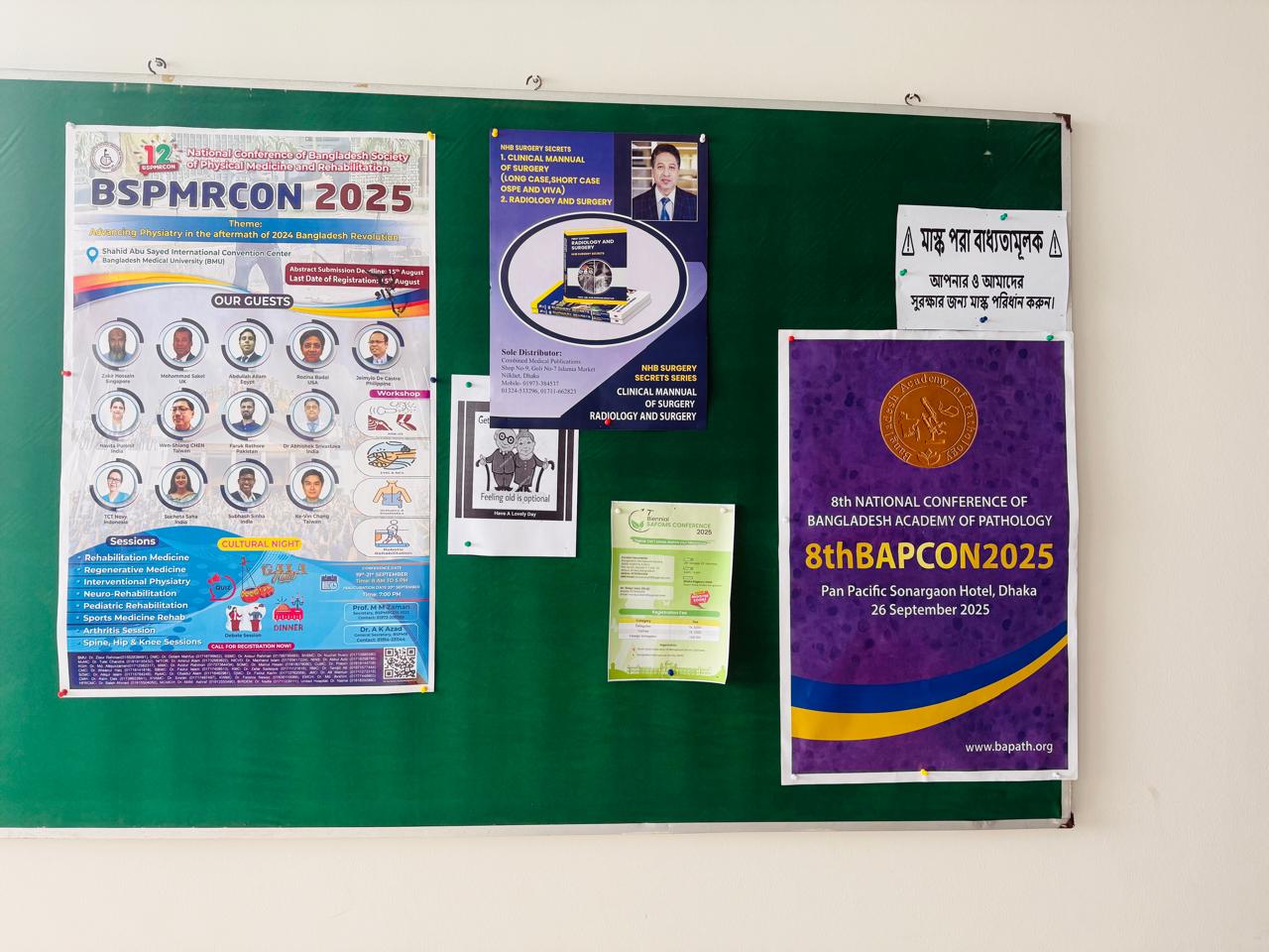


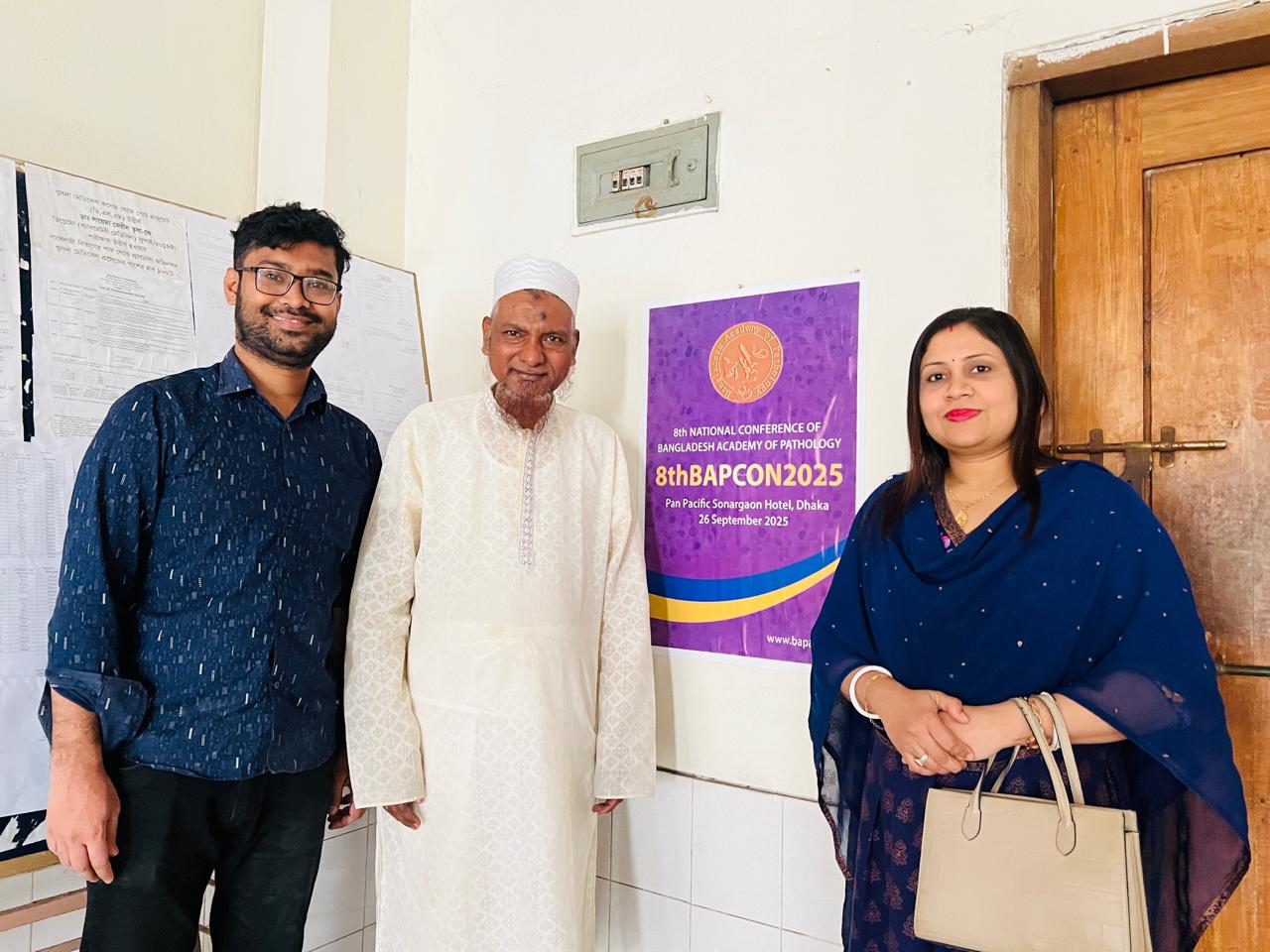


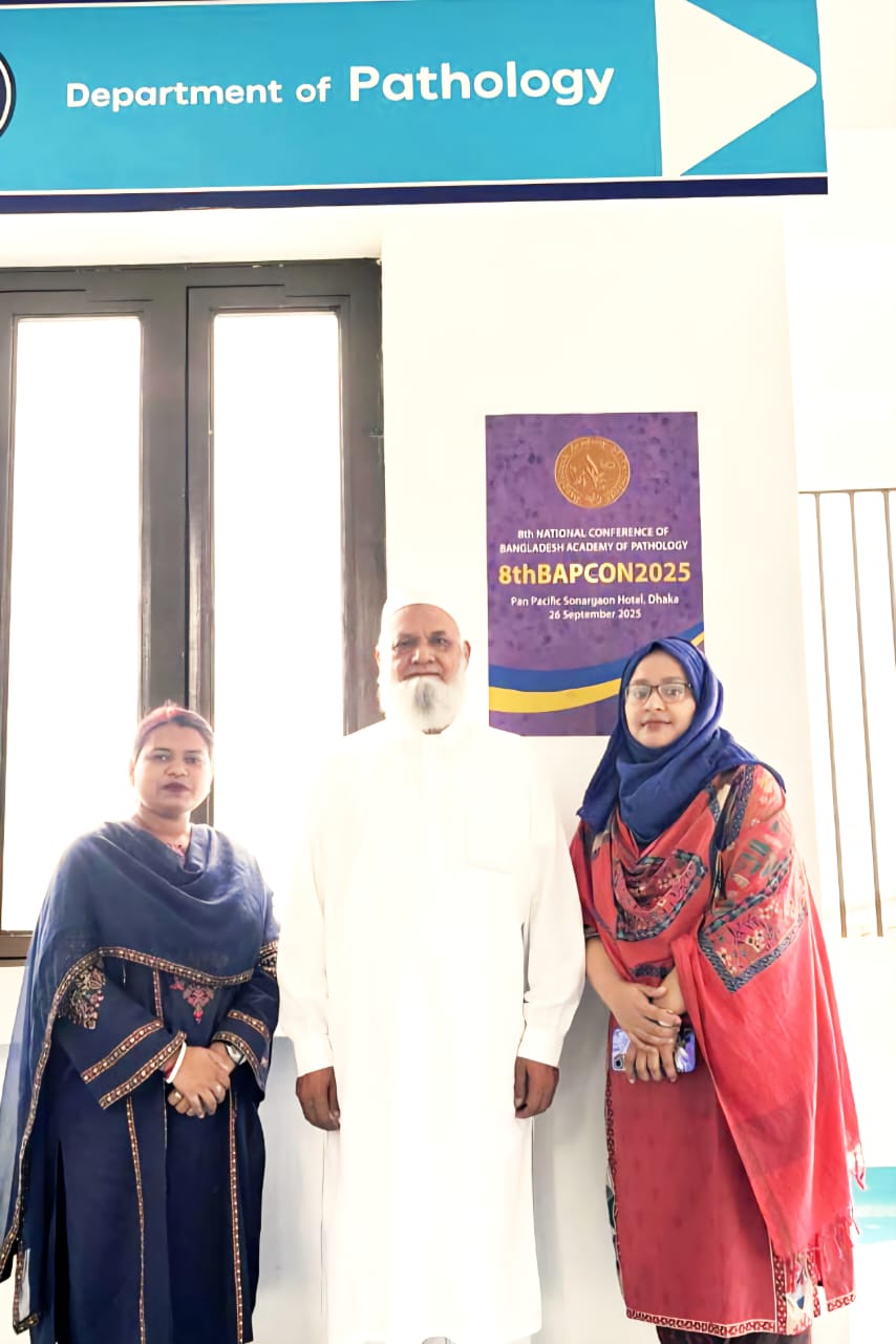


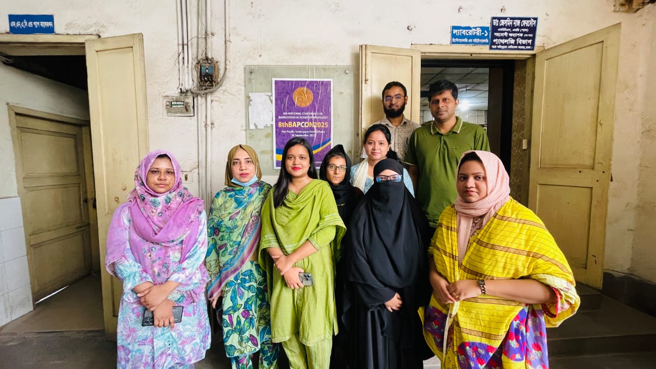


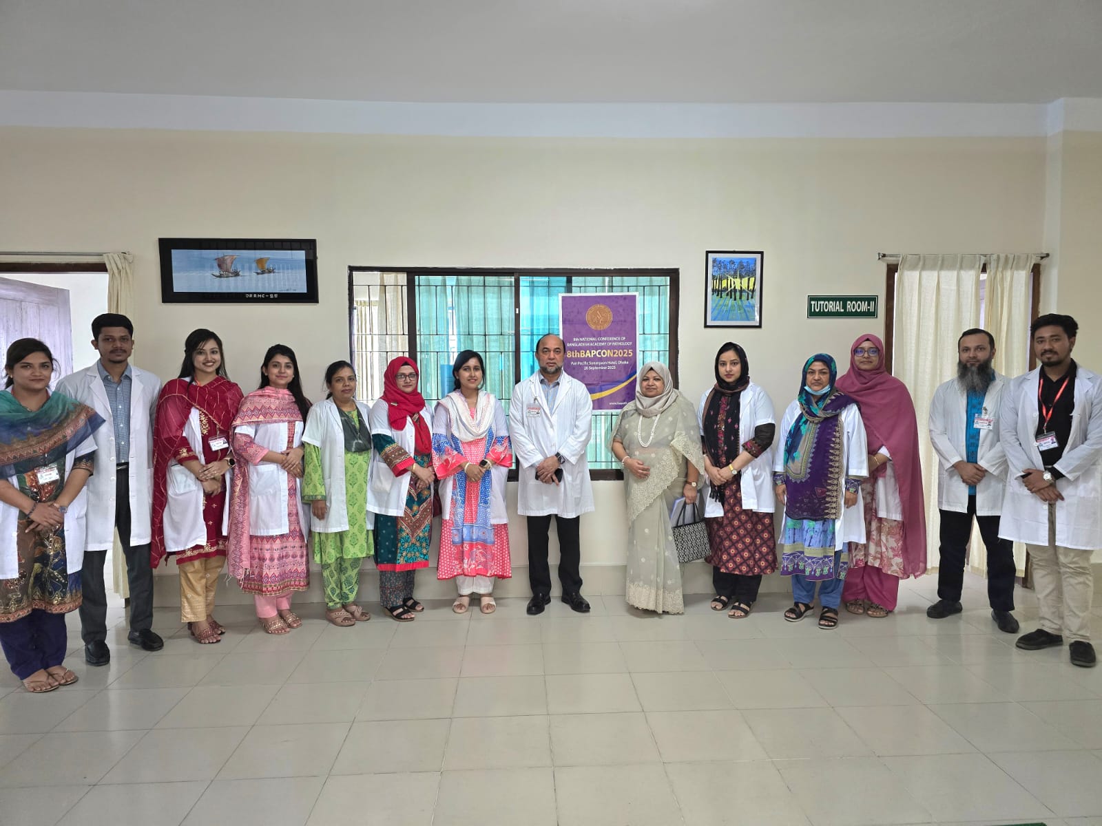


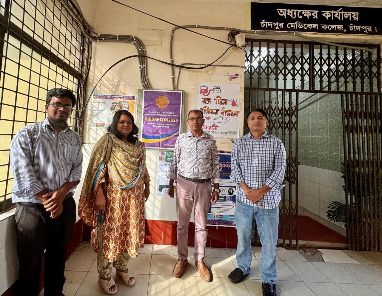
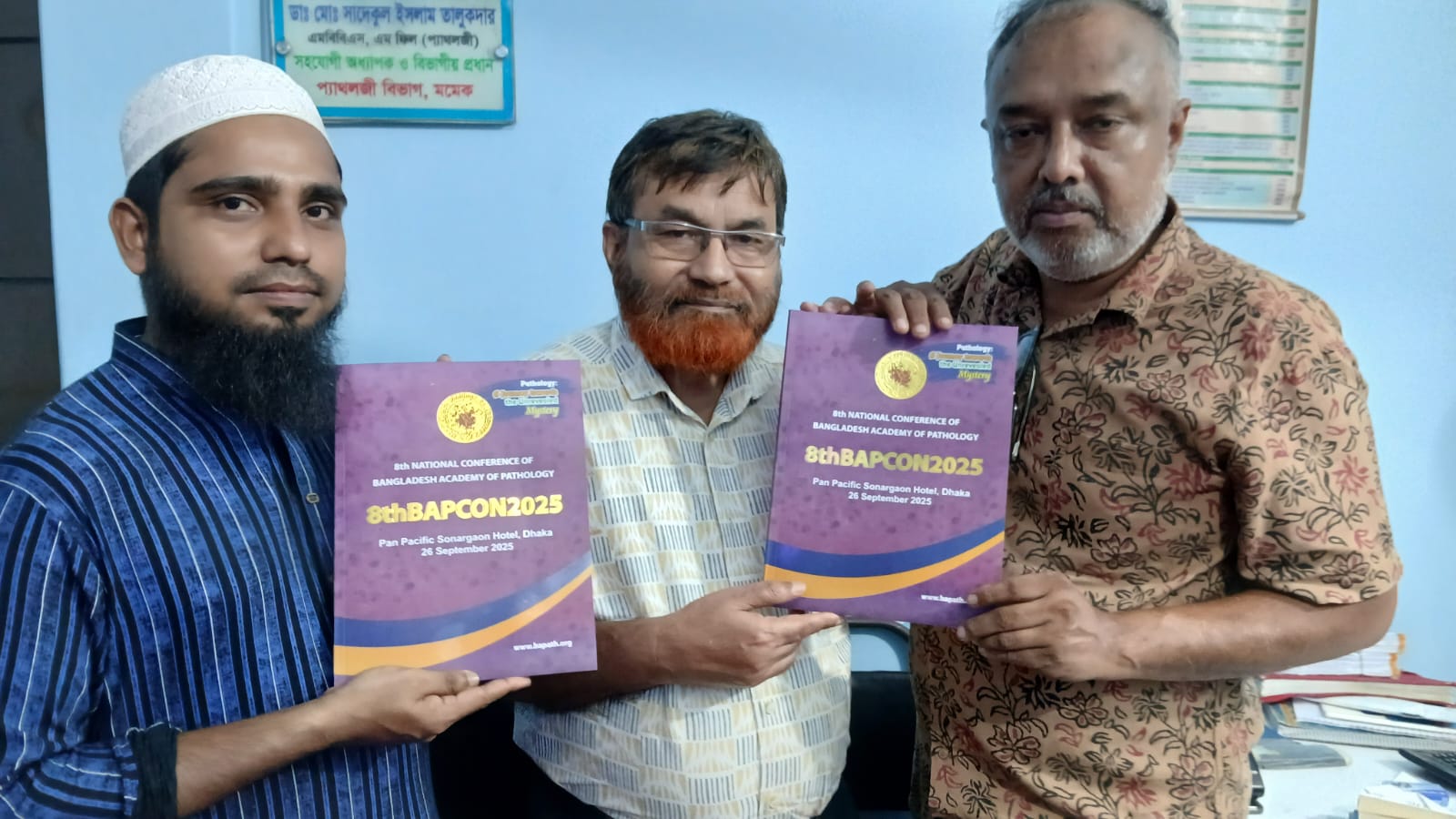


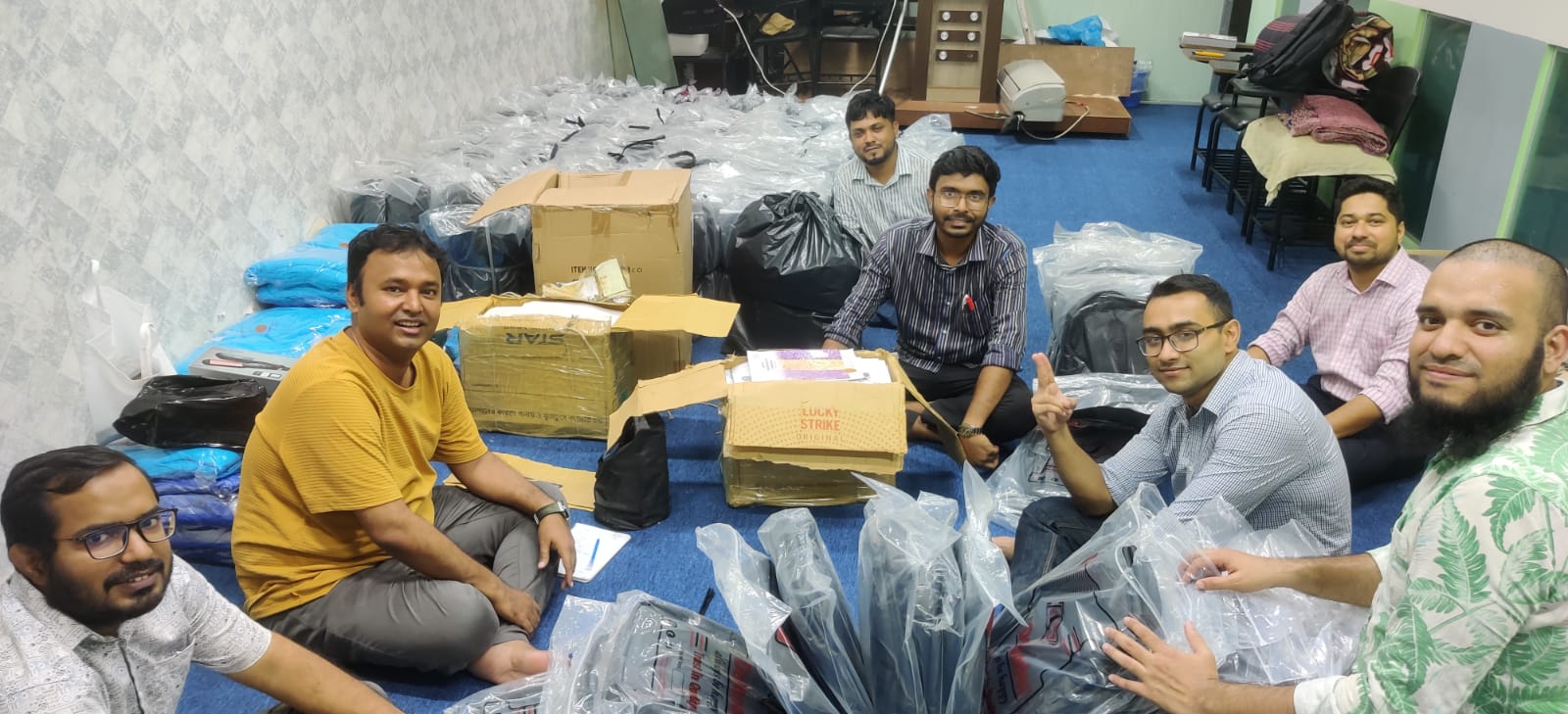


Conference Related Pictures Open
Conference Related Videos Open
Journal of Histopathology and Cytopathology, 2025 Jul; 9 (2)
Editorial
Digital Pathology: Prospects and Challenges
*Rahman DA,1 Akter S2
*For correspondence
Abstract: Not available
[Journal of Histopathology and Cytopathology, 2025 Jul; 9 (2):68-69]
DOI: https://www.doi.org/10.69950/jhc2025v9i2s1