jhc-2019-jul-v-3-n-2-The-Role-yasmin-i

The Role of Special Stains in Trephine Biopsy of Lymphoma

 *Yesmin I,1 Begum F,2 Yunus A,3 Kabir AL,4 Baqui MN5

 *Dr. Israt Yesmin, Specialist Pathology, Pathology and Laboratory Medicine, Square Hospital, Dhaka. badhanydr29@gmail.com

  1. Ferdousy Begum, Associate Professor, Department of Pathology, Bangabandhu Sheikh Mujib Medical University, Dhaka. ferdousy_begum2000@yahoo.com
  2. ABM Yunus, Professor & Chairman, Department of Haematology, Bangabandhu Sheikh Mujib Medical University, Dhaka. abmyunus.bsmmu@gmail.com
  3. Amin Lutful Kabir, Associate Professor, DEPT of Haematology, BSMMU Email aminlutful@gmail.com
  4. Muhammad Nazmul Baqui, Senior Lecturer, Faculty of Medicine, AIMST University, Kedah, Malaysia. doctornazmul@yahoo.com

*For correspondence

 Abstract

Background: Accurate diagnosis of lymphoma is always challenging to histopathologists. Aside from routine H& E stain special stains play an important role inreaching the final diagnosing of  lymphomacases. Here we have studied the use of these special stains in trephine biopsy material of lymphoma cases.
Methods: This descriptive cross sectional study was carried out at the Department of Pathology Bangabandhu Sheikh Mujib Medical University (BSMMU), Dhaka, from July 2012 to June 2014. Total 17 cases, which underwent trephine biopsy were included in this study. Different special stains and immunostains were done to reach the final diagnosis. Statistical analyses were performed to find out the usefulness of these special stains.
Results: Fourteenpatients were male and rests were female. Mean age of the patients was 41.8±12.5 years. IHC of trephine biopsy material aids in diagnosis of lymphomatous infiltration with further sub classification. While conventional H&E and giemsa stain revealed only 6(35.3%) cases with lymphomatous infiltration of bone marrow. Among these 14 cases, 10 (58.8%) cases were B-NHL, 3 (17.6%) cases were T-NHL and rest 1(5.9%) was lymphocyte-rich classical Hodgkin lymphoma.
Conclusion: It was found that, use of special stains and IHC of trephine biopsy material of lymphoma cases improved the diagnostic accuracy of lymphoma.

[Journal of Histopathology and Cytopathology, 2019 Jul; 3 (2):134-142]

Key words: Trephine biopsy

Introduction

Trephine biopsy of the bone marrow has wide application in clinical medicine. Its important use is in the evaluation of patients with various haematological disorders like malignant lymphoma, acute leukaemias, myeloproliferative disorders, myelodysplastic syndromes, metastatic tumor, granulomatous disorder, myelofibrosis, aplastic anaemia and plasma cell dyscrasias. Examination of the bone marrow aspirate and trephine biopsy is essential for the diagnosis of bone marrow disorders. The aspirate and trephine biopsy provide complimentary and useful information. It is recommended that both bone marrow aspirate and biopsy be routinely performed for correlation of representative findings.

The bone marrow trephines are particularly useful in identifying focal lesion that may be less apparent on aspirate smear alone. The bone marrow biopsy is also a common and important staging procedure that is performed routinely on patients with hematologic and non-hematologic malignancy. Pathomorphological examination of trephine biopsies of the bone marrow represents a standard method for the diagnosis and staging of hematological neoplasms and other disorders involving the bone marrow.  The use of special stains in bone marrow pathology should be determined following examination of the routinely stained biopsy and the patient’s clinical history.1 Although conventional morphology remains the gold standard for paraffin embedded BM trephines, IHC staining has become an integral part of the diagnostic and prognostic workup.2 Distinction of the most common type of B-NHL in the trephine biopsy can be difficult on morphological ground alone due to overlapping cytological features and distribution pattern and therefore require IHC and occasionally molecular data for primary diagnosis.2

Non Hodgkin lymphoma (NHL) composed of small B lymphocytes comprises the majority of lymphoma infiltrates in the bone marrow. Bone marrow involvement is seen in virtually 100% of B cell chronic lymphocytic leukemia (B-CLL) / small lymphocytic lymphoma (SLL), in almost all cases of lymphoplasmacytic lymphoma (LPL) and hairy cell leukemia (HCL), in 55-93% of mantle cell lymphoma (MCL), in 50-60% of follicular lymphoma (FL) and invariably in splenic marginal zone B cell lymphoma (SMZL). In nodal and extra nodal marginal zone B cell lymphoma the marrow involvement is found in 10-30% cases.2

The pattern of bone marrow involvement is one of the most important hints for diagnosis. Different patterns of bone marrow infiltration by NHLs are recognized, namely paratrabecular, randomly focal / intertrabecular / non paratrabecular, interstitial, diffuse and sinusoidal / intravascular. In some cases, multiple or mixed pattern are present in the same specimen. Although most infiltrates are easily recognizable in standard stains, sparse interstitial and sinusoidal involvement can be practically undetectable by conventional morphology and requires IHC to highlight the neoplastic population.2

Lymphoma demonstrate significant increased incidence rates throughout the world and causes mortality.3 Though uses of special histochemical stain in trephine biopsy is an established practice in histopathology, the application of immunohistochemistry (IHC) to diagnostic bone marrow trephine biopsy specimen is a relatively new practice.4 IHC is a powerful auxiliary technique for the diagnosis of hematologic disorder in bone marrow trephine biopsies.2 IHC reveals clear architectural features and estimates tumor cellularity in paraffin sections. By using IHC an accurate characterization and primary classification of lymphoid neoplasm is possible in the majority of cases and expands the diagnostic scope of bone marrow histology.2 The ability to define such subgroups is increasingly important as treatment is tailored not only to specific types of lymphoma but also on the basis of their prognostic group.5 Therefore the aim of this study was undertaken to see the role of special stains in bone marrow trephine biopsy in the diagnosis of lymphoma and to detect lymphoma of bone marrow and their typing.

Methods

This descriptive cross sectional study was carried out at the Department of Pathology Bangabandhu Sheikh Mujib Medical University (BSMMU), Dhaka, from July 2012 to June 2014. A total of 17 patients of different age and sex were selected from the department of Hematology BSMMU, Dhaka and other different institutes of Dhaka by using non probability sampling method. Patients who underwent trephine biopsy and histologically and clinically suspected as cases of lymphoma and cases accompanied by clinical information were included in this study. Exclusion criteria include patients who were not suspected as cases of lymphoma in histological diagnosis, specimen having severe crush artifact and specimen less than 1 cm in length. According to standard protocol the biopsy samples were taken from the posterior superior iliac spines under local anesthesia. Formalin fixed trephine biopsy sample were allowed to fix over night at room temperature in 10% buffered formalin fixative. Trephine biopsy samples collected from haematology department of BSMMU and other different institutes of Dhaka were decalcified in 10% formic acid for four hours followed by wash in running tap water for an hour.  Routine processing and paraffin embedding were done according to BSMMU standard tissue processing protocol in automated machines.  Paraffin blocks were made.  Subsequent tissue sections were analyzed by using routine H&E stain, periodic acid Schiff (PAS) stain, Giemsa stain and a primary histological diagnosis were made.

Sections with at least five well preserved marrow spaces were studied for cellularity, normal hematological elements, presence of infiltration if any, the extent, histologic pattern and morphology of infiltration and other secondary changes. The morphology and histologic pattern of infiltration were categorized as diffuse, interstitial, focal (non-paratrabecular), paratrabecular and mixed patterns. Diffuse pattern was defined when there was extensive replacement of the marrow elements, both hematopoietic tissue and fat, obscuring the marrow architecture.  Interstitial pattern was indicated when individual neoplastic cells were interspersed between hematopoietic cells and fat, focal, where nodular aggregates were seen separated by normal hematopoietic marrow, paratrabecular, when infiltrate aggregates were seen immediately adjacent to bony trabeculae.

2–3 μm thick sections were cut from the paraffin blocks for IHC. EnVision method was followed. The immunohistochemical staining procedure of all antibodies except TdT was performed in the IHC lab of BSMMU by following its staining protocol. Table I shows the IHC panel done for diagnosis.

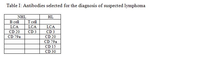
Results of immunostains were assessed by light microscopy using a 10× objective lens with subsequent use of 20 and 40 lenses for confirmation. Immunostains results were expressed as positive, weakly positive or negative. Cases where quantification of specific type of immunohistochemical markers positive cells are needed counted at x40 magnification, excluding cortical and trabecular bone, periosteal connective tissue, adipose tissue or areas of haemorrhage. The specific immunohistochemical marker positive cells were counted in five or more randomly selected fields and the number of positive cells was expressed as a percentage of total number of bone marrow nucleated cells.  Slides were reviewed by two senior histopathologists. Subsequently special histochemical stains and immunohistochemical finding was analyzed and final reports were issued. This was performed in the department of pathology of BSMMU, Dhaka.

 Results

Age, sex and clinical findings

It was observed that maximum case (35.3%) patients belonged to age grouped 31-40 years. The mean age was found 41.8±12.5 years with range from 20 to 70 years. Majority14 (82.4%) patients were male and 3(17.6%) were female indicating male predominance. Male and female ratio was 4.7:1. Out of 17 patients of the present study 13 patients had fever, 6 complained of weakness and 6 presented with hemorrhagic manifestation e.g. gum bleeding, cyanosis, bleeding spot etc. The other complains included weight loss, bone and joint pain, swelling of whole body, night sweat, pruritus, yellow discoloration of skin and sclera, neck swelling, oral ulcer etc. Anaemia was the most prevalent (88.2%) feature. Other signs included jaundice, oedema, cyanosis etc. Five (29.4%) patients had generalized lymphadenopathy, two (11.8%) had grouped palpable lymph node, one (5.9%) had bony tenderness and all had normal tonsil.

 Ultrasonographic findings

Among 17 cases USG reposts were not available in 5 cases. Two (11.8%) patients had splenomegaly, 2(11.8%) had hepatomegaly, 6(35.3%) had hepatosplenomegaly, 1(5.9%) had abdominal lymphadenopathy and USG findings were normal in 1(5.9%) cases.

Bone marrow examination

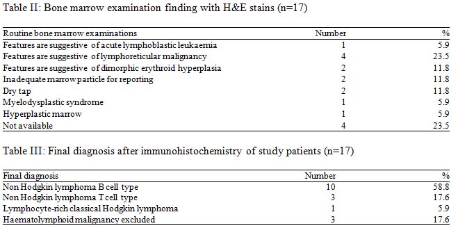
Comparison of Primary histological diagnosis with final diagnosis after IHC

Out of these 17 cases 14 cases as various types of lymphoma. In the present study, primary histological diagnosis on trephine biopsy was made as haematolymphoid malignancy unclassified in 11(57.1%) cases and NHL in 6(42.9%) cases. Fibrosis was not seen in any of the 17 cases.


Trephine biopsy samples of 17 cases were histologically analyzed with the help of H&E stain along with PAS and Giemsa stain. Five patients were finally diagnosed as NHL by primary histological diagnosis was made on the basis of different pattern of abnormal cellular infiltration in the bone marrow trephine biopsy.

Discussion

In this series it was observed that most of the patients with haematological disorder were in 4th decade and their mean age was 36.1±15.01 years. Male to female ratio was almost 3:1. Similarly study conducted by Chen et al. (2000) observed male to female ratio was 2.9:1.6 Similar observations regarding the male predominant were also made by Matsuo et al. (2003) and Lone & Naeem (2011).7 On the other hand Kumar et al. (2009) observed female predominant, where they found female to male ratio was almost 5:1.8

 The pattern of marrow involvement also differs to some extent in various NHL subtypes, for example paratrabecular infiltration is strongly associated with follicular lymphoma. A bone marrow biopsy performed in patients with low grade lymphoma sometime shows unexpected high grade transformation, which necessitates a different therapeutic approach.9

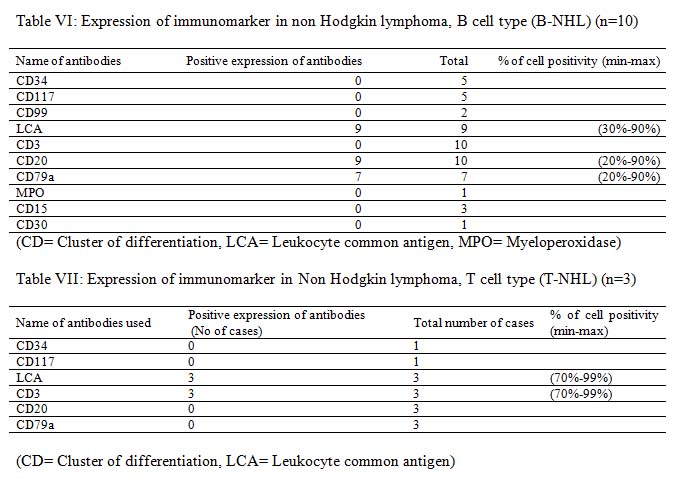
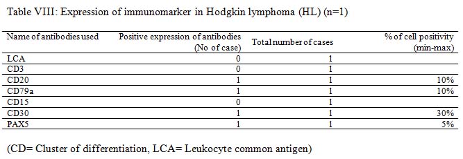
 Among initial 11 cases of haematolymphoid malignancy unclassified in trephine biopsy were finally seven of them were diagnosed as B-NHL and one case as Hodgkin lymphoma. The remaining three cases were finally excluded from haematolymphoid malignancy. This result indicates the importance of IHC in the diagnosis of haematolymphoid malignancy. The remaining six cases of NHL were further sub classified as B-NHL and T-NHL (Table IV).

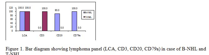
It is very much clear that trephine biopsy reports are superior as it could specify the diagnosis in most of the cases with the exception of cases of B-NHL and T-NHL. The latter cases were diagnosed only on the basis of CD3, CD20 and CD79a immunomarker. The incidence of bone marrow involvement in extra nodal MZL is lower (~20%) than in other low grade B-NHL, albeit recent studies described BM infiltration in up to 44% of cases.10

West et al. (2002) studied 61 cases (26 follicular lymphoma and 35 benign or atypical aggregates). They found that no single stain is sufficient for identification of neoplastic lymphoid aggregates. However, this distinction was made possible by using a panel of antibodies. The most useful antibodies were CD10, bcl-2, CD5, and CD20. Most benign or atypical aggregates do not express CD10 and CD23.11

Bone marrow specimens from 317 patients with non-Hodgkin’s lymphoma (NHL) obtained at initial staging was evaluated for the presence of lymphoma or benign lymphoid aggregates were studied by Conlan et al. (1990). They have found thirty-two percent (102 patients) had lymphoma in their bone marrow, and 9% had benign lymphoid aggregates. Bone marrow lymphoma was present in 39% of low-grade, 36% of intermediate-grade, and 18% of high-grade lymphomas. The bone marrow was involved in 25% of patients with diffuse large-cell or immunoblastic NHL. Bone marrow involvement did not affect survival of patients with low-grade NHL, but survival was significantly shorter (P<0.05) for patients with intermediate- and high-grade NHL with bone marrow involvement. Bone marrow involvement was equally common in B-cell and T-cell NHL (31% v 32%).12

Bone marrow involvement by lymphoma is considered a systemic dissemination of the disease arising elsewhere, although some tumors may arise primarily in the bone marrow microenvironment 13. IHC is important for lymphoma in particular those with heterogeneous histology to establish accurate diagnosis and adequate therapy14.

Bone marrow aspiration and bone marrow biopsy are important diagnostic procedures for diagnosis of hematological, non-hematological malignancies and other diseases. These procedures are also valuable for follow up of patients undergoing chemotherapy, bone marrow transplantation and other forms of medical treatment. In the study conducted by Toi et al. (2010),  comparative evaluation of all such bone marrow aspiration (BMA) and bone marrow biopst (BMB), to see the complementary role of both the procedures, to study the advantages and disadvantages of both the procedures done simultaneously. There was 61.25% positive correlation between BMA and BMB in the study15.

Conclusion

It can be concluded that correct diagnosis can be made in majority of cases with the application of PAS, Giemsa and limited number of immunomarker. Correct and relevant clinical information along with appropriate special stain of bone marrow trephine biopsy improves the diagnostic accuracy of lymphoma cases.

 References

  1. Brunning RD, Arber DA, 2011. Bone marrow. In J, Rosai Rosai and Ackerman’s Surgical Pathology, Tenth edition, Mosby Elsevier Inc, pp. 1927-2012.
  2. Kremer M, Quintanilla-Martínez L, Nährig J, von Schilling C, Fend F. Immunohistochemistry in bone marrow pathology: a useful adjunct for morphologic diagnosis. Virchows Arch Int J Pathol. 2005 Dec;447(6):920–37.
  3. Tahmasby B, Marnani AB, Maleki M, Barouni M, Mousavi SH, Naseriyan B, et al. Blood malignancies in Mazandaran province of Iran. Asian Pac J Cancer Prev APJCP. 2013;14(2):1053–6.
  4. Olsen RJ, Chang C-C, Herrick JL, Zu Y, Ehsan A. Acute leukemia immunohistochemistry: a systematic diagnostic approach. Arch Pathol Lab Med. 2008 Mar;132(3):462–75.
  5. Guidelines on the management of acute myeloid leukaemia in adults – – 2006 – British Journal of Haematology – Wiley Online Library [Internet]. [cited 2019 Sep 30]. Available from: https://onlinelibrary.wiley.com/doi/full/10.1111/j.1365-2141.2006.06314.x
  6. Clinicopathological studies on bone marrow involvement of non-Hodgkin’s lymphoma [Internet]. ResearchGate. [cited 2019 Sep 30]. Available from: https://www.researchgate.net/publication/12095478_Clinicopathological_studies_on_bone_marrow_involvement_of_non-Hodgkin’s_lymphoma
  7. Matsuo T, Kuriyama K, Miyazaki Y, Yoshida S, Tomonaga M, Emi N, et al. The percentage of myeloperoxidase-positive blast cells is a strong independent prognostic factor in acute myeloid leukemia, even in the patients with normal karyotype. Leukemia. 2003 Aug;17(8):1538–43.
  8. Kumar S, Rau AR, Naik R, Kini H, Mathai AM, Pai MR, et al. Bone marrow biopsy in non-Hodgkin lymphoma: a morphological study. Indian J Pathol Microbiol. 2009 Sep;52(3):332–8.
  9. Juneja SK, Wolf MM, Cooper IA. Value of bilateral bone marrow biopsy specimens in non-Hodgkin’s lymphoma. J Clin Pathol. 1990 Aug;43(8):630–2.
  10. Feugier P, De March AK, Lesesve JF, Monhoven N, Dorvaux V, Braun F, et al. Intravascular bone marrow accumulation in persistent polyclonal lymphocytosis: a misleading feature for B-cell neoplasm. Mod Pathol Off J U S Can Acad Pathol Inc. 2004 Sep;17(9):1087–96.
  11. West RB, Warnke RA, Natkunam Y. The usefulness of immunohistochemistry in the diagnosis of follicular lymphoma in bone marrow biopsy specimens. Am J Clin Pathol. 2002 Apr;117(4):636–43.
  12. Conlan MG, Bast M, Armitage JO, Weisenburger DD. Bone marrow involvement by non-Hodgkin’s lymphoma: the clinical significance of morphologic discordance between the lymph node and bone marrow. Nebraska Lymphoma Study Group. J Clin Oncol Off J Am Soc Clin Oncol. 1990 Jul;8(7):1163–72.
  13. Primary bone marrow lymphoma: an uncommon extranodal presentation of aggressive non-hodgkin lymphomas. – PubMed – NCBI [Internet]. [cited 2019 Sep 30]. Available from: https://www.ncbi.nlm.nih.gov/pubmed/22251943
  14. Bone marrow aspiration before bone marrow core biopsy using the same bone marrow biopsy needle: a good or bad practice? [Internet]. [cited 2019 Sep 30]. Available from: https://www.ncbi.nlm.nih.gov/pmc/articles/PMC1860622/
  15. Toi PC, Varghese RG, Rai R. Comparative evaluation of simultaneous bone marrow aspiration and bone marrow biopsy: an institutional experience. Indian J Hematol Blood Transfus Off J Indian Soc Hematol Blood Transfus. 2010 Jun;26(2):41–4.

jhc-2019-jul-v-3-n-2-study-ansari-m

Study of Ovarian Tumours: Histomorphological Types

 *Ansari M,1  Khan AH,2 Hossain S,3 Khanom F4

  1. *Dr. MAS Ansari, Associate Professor of Pathology, Sylhet Women’s Medical College, Sylhet. drmasansari@gmail.com
  2. Professor Dr. Amjad Hossain Khan, Professor of Pathology, Sylhet Women’s Medical College, Sylhet.
  3. Sabbir Hossain, Associate Professor of Pathology, North East Medical College, North East Medical College, Sylhet.
  4. Farida Khanom, Medical Officer of Gynae & Obs, Jalalabad Ragib-Rabeya Medical Colleg, Sylhet.

 *For correspondence

 Abstract

Background: Ovarian tumour accounts for 3% of total cancer in females and is the 5th most common form of cancer related death in females with a varied clinical, morphological and histological features.

Objectives: The present  study was carreid out with an aim to find out  the frequency, age distribution and the histopathological  patterns of ovarian tumours.
Methods: Retrospective  study was done during the period  of 3 years comprised of 125 ovarian tumours diagnosed in the Department of Pathology, Sylhet Women’s Medical College, Sylhet. After thorough gross examination, representative bits were routinely processed and stained with H & E. Tumours were classified as per WHO classification.
Results: Out of 125 cases studied, majority were benign tumours (88%), followed by malignant (10.4%) and borderline tumour (1.6%). Age ranged from 11-70 years. Epithelial tumours were most common (76%), followed by germ cell tumours (16%), sex cord stromal tumours (7.2%) and metastatic ovarian tumours (0.8%). Serous cystadenoma was the commonest benign tumour and serous cyst adenocarcinoma was the commonest malignant ovarian tumour.
Conclusion: The prognosis and varying therapeutic strategies of ovarian tumours necessitate an accurate pathological evaluation. Although newer techniques like IHC and molecular analysis have made the diagnosis easier and more precise, in the institutes with provision of limited resources, histopathological study is still the gold standard in diagnosing most of these tumours.

 [Journal of Histopathology and Cytopathology, 2019 Jul; 3 (2):125-133]

Key words: Ovarian tumour, Histopathology

 Introduction

Ovarian tumours include complex, wide spectrum of neoplasms involving a variety of histological patterns ranging from epithelial tissues, connective tissues, specialized hormone secreting germinal and embryonal cells.1

The poor survival is due to the fact that they do not clinically manifest early and approximately 60-70% of the neoplasms present as either stage III or stage IV.1,3,4 Benign ovarian cysts are the commonest constituting about 80% of ovarian tumours and mostly occur in young women between the ages of 20-40 years. Borderline tumours occur at slightly older ages whereas the malignant tumours are common in older women between the ages of 40-65 years.1,4 Metastatic tumours subsequently involve the ovaries and mimic primary ovarian neoplasia. Approximately, 7% of lesions presenting clinically as primary ovarian tumours are of metastatic origin.5, 6 It is important to determine the histological pattern of ovarian tumour to achieve the optimum treatment response as prognosis depends on the degree of differentiation.2,3,7

Thus present study was undertaken to analyse the frequency of various histological subtypes, age distribution pattern and the  histopathological pattern of ovarian tumours.

 Methods

This retrospective  study was conducted for a period of 3 years (January2015 – December 2018) at Sylhet women’s Medical College, Sylhet.

Specimens sent in 10% formalin were routinely processed with paraffin embedding after adequate fixation. Paraffin sections and slides from fresh blocks and the retrieved blocks were stained with H & E. The slides were then reviewed microscopically in detail and tumours were classified according to the WHO classification of ovarian tumours.

Inclusion criteria

All histologically proven both primary and secondary ovarian tumours.

Exclusion criteria

Non- neoplastic ovarian lesions of study period.

Results

A total number of 125 cases were studied. Among these, majority were primary ovarian tumours (124; 99.2%), while one was metastatic ovarian tumours (1; 0.8 %). Of them 69 cases were benign (88%), 15 cases were malignant (10.4%) and rest 2 cases (1.6%) were borderline. Epithelial tumours (ET) were the most common histological type (95;76%), followed by Germ cell tumours (GCT) (20;16%) and sex cord stromal tumour (SST 9;7.2%; Table I).

Age Distribution

Majority of the tumours occurred in the reproductive age group (57; 45.6%) followed by 41-50 years of age group (26; 20.8%).Youngest patient was 13 years of age and older patient was about 68 years. Epithelial tumours and Sex cord stromal tumours had its peak between 31 to 40 years, whereas Germ cell tumours showed a peak in 21 to 30 years. Metastatic tumoursshowed  distribution between 61-70 years of age group.

 Gross Features

Most of the cases in this study were unilateral (117; 93.6%) and few were bilateral (8; 6.4%). Out of the total 95 Epithelial tumours, 76 were cystic in nature (80%), followed by those with cystic to solid in consistency (18; 18.9%) and solid (1; 1.05%), whereas most of Germ cell tumours were cystic in nature (16; 72.7%), followed by solid in consistency (5; 22.7%). Majority of sex cord stromal tumours (5; 71.4%) and all of the metastatic tumours were partly solid to cystic in consistency.

 Microscopy

The most common benign tumour was Serous cystadenoma (54; 43.2%), followed by Mucinous cystadenoma (14; 11.2%) and Mature cystic teratoma (16; 12.8%). Serous cyst adenocarcinoma was the most common malignant tumour (6; 4.8%), followed by Adult granulosa cell tumour (3; 2.4%). Borderline serous tumour were two in numbers. There was one case of metastatic ovarian tumours, was Krukenbergtumour. (Table II).

Discussion

Ovarian tumours are one of the major health problems and their diagnosis can be difficult due to variety of pathologic conditions affecting the ovaries. Thus knowledge of morphology and age-specific characteristics can help refine the diagnosis.8, 9

In the present study of 125 ovarian tumours, Primary ovarian neoplasms (99.2%) were the most common tumours of all ovarian tumours. Similar observations were made by Bhagyalakshmi A et al.10 (98.5%) and Vaddatti T et al.3 (98.9%). Most of the tumours belonged to Epithelial tumour category (76%) which was comparable to the results seen by Singh S et al.11 (69.17%), Krishna M & Maurya G12 (77.7%) and Badge S et al.13 (77%). GCT and SST accounted for 16% and 7.2%, respectively in our study compared to 42.2% and 3.1% reported in the others.14

In the present study, majority of the tumours were benign (88%) followed by malignant tumours (10.4%) and rest was borderline (1.6%). Findings of the present study correlated well with the studies of various authors.However, in this study the frequency of malignant tumours (10.4%) was little less than the study of Couto F et al.15 (40%).

In the present study, the patient’s age ranged from 11 years to 70 years and this was supported by the study done by Swamy GGEt al.16 where the youngest patient was 13 years old and the oldest was 68 years old. The majority of ovarian tumours (60.7%) were seen in the age group of 21-40 years, which was consistent with the study done by Kuladeepa A VK et al.17 (58.9%) and Pilli GS et al.18 (58.3%) .

In our study, majority of the tumours were unilateral (97.1%) and least were bilateral (2.9%) which were consistent with the findings of  VaddattiT et al.3, Jindal U8 and Prabhakar BR and Maingi K.19

In the present study, majority of the tumours were cystic (72%), followed by partly cystic & solid (18.4%) and few were solid (9.6%). These findings were comparable with the findings of Misra RK et al.20 where most of the tumours were cystic (78.2%) and few were solid (4.1%), whereas Couto F et al.15 have found 10.2% solid tumours which was close to our findings. Benign epithelial tumors were the commonest type (88.4%), followed by malignant epithelial tumors (9.47%) and the rest was borderline (2.10%). These findings were almost similar as observed in the study conducted by Kuladeepa A VK et al.17) Sharma I et al.21

In this study, maximum number of epithelial tumors (78%) were noted in 31-50 years age group. All malignant tumours were seen in the cases above 50 years of age. The results corroborated with the various studies done by Tushar K et al. (2005),22 Jha R & Karki S14 and Jindal U.8 Among the histomorphological types of epithelial tumors, Seroustumours (73.6%) were the most common, followed by Mucinous tumours (16.8%), Seromucinoustumours (8.42%) and least common were endometrioid tumours (1.05%). Similar results were seen by Krishna M & Maurya G12 andSarkar R.23 Serous cystadenoma was the commonest ET (53.3%) followed by Mucinous cystadenoma (21.7%) and serous cyst adenocarcinoma (8.4%). Similar results were reported by various authors in their studies.

In the present study, majority of the  germ cell tumours were benign (81.8%) and include mature cystic teratoma and Struma ovarii. These results were closer to the findings of Agrawal P et al.24 and Verma K & Bhatia A25 with 77.7% and 83.5% respectively. Malignant tumours include Dysgerminoma and Embryonal carcinoma. Germ cell tumours showed maximum cases below 35 years of age, with a gradual decline in 31-40 years age group and they were found to be uncommon after the age of 60 years. These findings were consistent with the studies of Jha R & Karki S14 and Agrawal P et al.24

In this study, majority of the sex cord stromal tumours were benign (57.1%) and the results were not similar to the findings of Rao KN et al.26 (55.6%) and were lower when compared to the findings of Jindal U8 (75%). On the contrary study of Badge SAEt al.13 (66.7%) showed mostly benign sex cord stromal tumours against malignant. The age range of sex cord stromal  tumours in our study was 41-70 years whereas Bhagyalakshmi A Et al.10 recorded age range of 21-70 years in their study. Adult granulosa cell tumour which is potentially malignant tumour occurred in 2.4% of all ovarian tumours. The frequency was consistent with the findings of Gupta SC et al.27 and Pilli GS et al.18 who recorded it as 4.4% and 3.54% respectively.

Our study showed one case of secondary tumours and constituted 0.8% of all ovarian tumours. This was lower when compared to the findings of Badge SA Et al.13  (10.9%) but was almost similar to the studies of Gupta N et al.28 (2%), Jha R & Karki S14 (2.4%) and Bhagyalakshmi A Et al.10 (1.5%). The former when compared with the studies of Misra RK et al.20 (1.07%), Prabhakar BR and Maingi K19 (1.57%) and Couto F et al.15 (1.46%) showed a good correlation.

 Conclusion

Ovarian tumour is a silent menace that presents as a tremendous clinical challenge to gynaecologist, medical oncologist and radiotherapists. Emergence of borderline tumours with prognostic difference from the benign and malignant counterparts, has added a wing to research in the field of ovarian tumours. Accurate diagnosis of ovarian tumours can be rendered in almost all of cases by correlating the clinical presentation, radiographic appearance and histomorphological features, which remains the gold standard. Even then, in the modern era by the application of specialised methods like special stains, IHC markers, ultrastructural studies and cytogenetics, there is a vast scope for reaching specific & reliable diagnosis of difficult dilemmatic cases of ovarian tumours, by which the therapeutic and prognostic implications could be modified.

References

  1. Pradhan A, Sinha AK, Upreti D. Histopathological patterns of ovarian tumors at BPKIHS. Health Renaissance. 2012; 10(2):87-97.
  2. Malli M, Vyash B, Gupta S, Desai H. A Histological study of ovarian tumors in different age groups.Int J Med Sci Public Health. 2014; 3(3):338-341.
  3. Vaddatti T, Reddy ES, Vahini G. Study of morphological patterns of ovarian neoplasms. IOSR Journal of Dental and Medical Sciences. 2013; 10(6):11-16.
  4. Ellenson LH, Piorg EC. The Female Genital Tract.Kumar V, Abbas AK, Aster JC. In: Robbins and Cotran Pathological Basis of Diseases. 9th edn: Elseiver, 2014; 2:1022-1034.
  5. Prat J, Morice P. Secondary tumours of ovary. Tavassoli FA, Devilee P. In: WHO classifications of tumours: Pathology of the breast and female genital organs. 3th edn. IARC Press: Lycon, 2003, 193.
  6. Rosai J. Female reproductive system- Ovary. In: Rosai and Ackerman’s Surgical Pathology. 10th edn. Missouri: Elsevier, 2012; 2:1553-1609.
  7. Sohail I, Hayat Z, Saeed S. A comparative analysis of frequency and patterns of ovarian tumours at a tertiary care hospital between two different study periods 2002-2009. J Postgrad Med Inst. 2012; 26(2):196-200.
  8. Jindal U. Pattern of ovarian neoplasm in rural population: A five year study from tertiary care hospital. Journal of evolution of medical and dental sciences. 2014; 3(8):2033-2039.
  9. Mondal SK, Banyopadhya R, Nag DR, Roychowdhury S, Mondal PK, Sinha SK. Et al. Histologic pattern, bilaterality and clinical evaluation of 957 ovarian neoplasms: A 10-year study in a tertiary hospital of eastern India. J Can Res Ther. 2011; 7(4):433-437.
  10. Bhagyalakshmi A, Sreelekha A, Sridevi S, Chandralekha J, Parvathi G, Vekatalakshmi A. Et al. Prospective study of histopathological patterns of ovarian tumours in a tertiary care centre. Int J Res Med Sci. 2014; 2(2):448-456.
  11. Singh S, Saxena V, Khatri SL, Gupta S, Garewal J, Dubey K. Et al. Histopathological evaluation of ovarian tumors. Imperial Journal of Interdisciplinary Research. 2016; 2(4):435-439.
  12. Krsihna M, Maurya G. Pattern of ovarian tumors and their age distribution in Kangra Valley Himachal Pradesh. Journal of Evolution of Medical and Dental Sciences. 2015; 4(61):10602-10608.
  13. Badge SA, Gosavi AV, Sulhyan KR. Histopathological study of ovarian tumors. Indian Medical Gazette, 2013; 345-351.
  14. Jha R, Karki S. Histological pattern of ovarian tumors and their age distribution. Nepal Med Coll J. 2008; 10(2):81-85.
  15. Couto F, Naolkarni NS, Rebello MJ. Ovarian tumours in Goa: A clinic pathological study of ovarian tumours. J ObstetGynaecol of India. 1993; 43(3):408-12.
  16. Swamy GG, Saryanarayan N. Clinico pathological analysis of ovarian tumors – A study on five years samples. Nepal Med Coll J. 2010; 12(4):221-223.
  17. Kuladeepa AVK, Muddegowda PH, Lingegowda JB, Doddikoppad MM, Basavaraja PK, Hiremath SS Et al. Histomorphological study of 134 primary ovarian tumors.Adv Lab Med Int. 2011; 1(4):69- 82.
  18. Pilli GS, Suneeta KP, Dhaded AV, Yenni VV. Ovarian tumours: A study of 282 cases. J Indian Med Assoc, 2002; 100:423-424.
  19. Prabhakar BR, Maingi K. Ovarian tumors – prevalence in Punjab. Indian J PatholMicrobiol, 1989; 32:276-81.
  20. Misra RK, Sharma SP, Gupta V, Gaur R, Mishra SD. International Journal of Medical and Health Research ,Pattern of ovarian neoplasm in eastern UP. J ObstetGnecol. 1991; 41(2):242-6.
  21. Sharma I, Sarma U, Dutta UC. Pathology of ovarian tumour- A hospital based study. International Journal of Medical science and Clinical Invention. 2014; 1(6):284-286.
  22. Tushar K, Asaranti K, C MP. Intra-operative cytology of ovarian tumours.J ObstetGynecol India. 2005; 55(4):345-349.
  23. Sarkar R. Ovarian neoplasms – A 14 years study. J ObstetGynecol India, 1996; 46:156-160.
  24. Agrawal P, Kulkarni DG, Chakrabati PR, Chourasia S, Dixit M, Gupta K Et al. Clinico pathological spectrum of ovarian tumors: a 5 year experience in a tertiary health care center. Journal of Basic and Clinical Reproductive Sciences. 2015; 4(2):90-96.
  25. Verma K, Bhatia A. Ovarian neoplasms: A study of 403 tumors. J ObstGynecol India.1981; 31:406-411.
  26. Rao KN, Koteswari M, Devi CP, Sailabala G, Katta R. Morphological study of ovarian tumors with special reference to germ cell tumors. IOSR Journal of Dental and Medical Sciences. 2015; 14(1):55-60.
  27. Gupta SC, Singh PA, Mehrotra TN, Agarwal R, A clinic pathological study of ovarian tumours: Indian J PatholMicrobiol. 1986; 29(4):354-62.

28. Gupta N, Bisht D, Agarwal AK, Sharma VK. Retrospective and prospective study of ovarian tumours and tumour-like lesions.Indian J PatholMicrobiol. 2007; 50(3):525-7.

jhc-2019-jul-v-3-n-2-ultrasound-ahmed-nu

Ultrasound Guided Fine Needle Aspiration Cytology and Histopathology in the Diagnosis of Ovarian Mass

 *Ahmed NU,1 Saha NK,2 Bhowmik DK,3 Mazumder AR,4 Shariar S,5 Hira AD,6 Keya SA7

  1. *Dr. Nasir Uddin Ahmed, Assistant Professor, Department of Pathology, Faridpur Medical College. nasirdr32@yahoo.com
  2. Naba Kumar Saha, Professor, Department of Pathology, Sylhet MAG Osmani Medical College.
  3. Dilip Kumar Bhowmik, Professor, Department of Gynaecology & Obstetrics, Sylhet MAG Osmani Medical College.
  4. Ashikur Rahman Mazumder, Professor, Department of Radiology & Imaging, Sylhet MAG Osmani Medical College.
  5. Sakib Shariar, Pathologist, Railway General Hospital, Dhaka.
  6. Ananda Dyuti Hira, Pathologist, Department of Pathology, Khulna Medical College.
  7. Shamim Ara Keya, Lecturer, Department of Pathology, Shaheed Suhrawardy Medical College, Dhaka.

* For correspondence

 Abstract

Objective: The study was designed to evaluate the efficacy of ultrasound guided fine needle aspiration cytology in the diagnosis of ovarian mass and to correlate with histopathological diagnosis.
Methods: This  cross sectional study was done on 60 patients. FNA smears were prepared before surgical procedure. Histopathological examination was done by collection of surgical specimens. Cytomorphological findings were noted and correlated with histomorphological diagnoses.
Results: The age of patients ranged from 16 to 70 years with a mean of 36 years. Study showed 59 (98.33%) were unilateral and 1(1.67%) tumor was bilateral. Among 60 histopathologically diagnosed cases, 7(11.67%) were non-neoplastic cysts, 40 (66.67%) were benign tumors, 2(3.33%) were borderline tumors, 10(16.67%) were malignant tumors and 1 (1.66 %) was inflammatory lesion. All hemorrhagic cysts and cystic lesions were considered benign because they were cytologically negative for malignancy. Concordant diagnosis between cytology and histopathology was in 57 (95%) cases.
Conclusion: USG guided FNAC showed a relatively safe, quick, cost effective and patient compliant procedure with minimal morbidity.

[Journal of Histopathology and Cytopathology, 2019 Jul; 3 (2):117-124]

 Key words: Ultrasound Guidance, Fine Needle Aspiration Cytology, Histopathology, Ovarian Mass.

 Introduction

Tumors of the ovary are incredibly varied. This multiplicity is due to the presence of three cell types in the normal ovary: the multipotent surface (coelomic) epithelium, the totipotent germ cells, and the sex cord–stromal cells, each of which gives rise to a number of different tumors.1 The clinicopathological evaluation of ovarian masses is a challenging field. Difficulty in gaining access to the tumor site is itself a major obstacle and the wide spectrum of lesions present an enigmatic picture to the pathologist. Although histopathology remains the gold standard, in recent times image guided aspiration is being increasingly used as a rapid, inexpensive and efficient method for the pre-surgical diagnosis of ovarian masses as well as planning and evaluation of treatment.2 Historically, gynaecologists are hesitant to aspirate ovarian masses in view of the possibility of seeding of an early stage ovarian cancer. The magnitude of risk of such a procedure is unknown and not substantiated by convincing evidence. It is rather overestimated and has not been pathologically confirmed.3

 However, a lot of studies have clearly documented that risk of tumor spreading by needle tract is negligible in comparison to the potential benefits of this simple, quick and effective modality of diagnosis.4 With sonographic support, any structure visualized radiologically can usually be reached precisely in any desired plane, thus increasing the cytological yield. Nevertheless, as with any technique, image-guided FNAC has its short comings; false-negative results are usually due to failure of the needle to enter the mass and failure to sample representative areas.5 In the preoperative diagnosis of ovarian masses, acellular fluid should not be considered non-diagnostic, because it represents benign cysts in a majority of the cases. Literature search failed to trace any study to see the effectiveness of USG guided FNAC in comparison with histopathology in the diagnosis of ovarian masses in our country. With this background, the study was designed to evaluate the efficacy of ultrasound guided fine needle aspiration cytology in the diagnosis of ovarian mass and to correlate with histopathological diagnosis.

 Methods

The cross sectional study was carried out in the Department of Pathology, Sylhet MAG Osmani Medical College, Sylhet in collaboration with the Department of Obstetrics and Gynaecology and Department of Radiology and Imaging, Sylhet MAG Osmani Medical College Hospital, Sylhet from 1st July 2014 to 30th June 2015. All patients with ovarian masses attended the inpatient department of Obstetrics & Gynaecology were considered as the target population and those who fulfilled the inclusion and exclusion criteria were considered as study population. Patient of all ages with ovarian mass were included in this study and patients with hemorrhagic diathesis, those who refused to undergo  USG guided FNAC and surgery, diagnosed cases under treatment and ovarian mass with pregnancy were excluded from this study. Aspirations were done by expert radiologist in radiology and imaging department. The tumors were located by USG and needle was inserted into the lesion without negative pressure. The content of the needle was expelled on to a glass slide and then allowed the aspirated material to spread by gently pulling apart two slides in opposite direction. The smeared slides were promptly dropped in 95% ethyl alcohol for fixation and kept for at least 30 minutes. This smears were then stained according to Papanicolaou method. The cytopathological examination of the stained slides was carried out on the same day or following day. Surgical specimens were collected from all patients in whom USG guided FNAC were done. All the specimens were preserved in 10% formalin. Routine tissue processing with paraffin impregnation was done and stained with hematoxylin and eosin. The diagnoses of ovarian tumors were done according to the cytopathological and histopathological findings and then cytopathological diagnoses were compared with the histopathological diagnoses.

After meticulous checking, all the relevant collected data were compiled first on a master table. Then the data were organized by using scientific calculator and standard computer based statistical software SPSS-21. Percentages were calculated to find out the proportion of the findings. The results were presented in tables, graphs and diagrams. The efficacy of USG guided FNA cytology for the diagnosis of ovarian mass was determined by calculating sensitivity, specificity, positive predictive values, negative predictive values and accuracy.

 Results

Total 62 cases were selected considering inclusion and exclusion criteria. Tissue for histopathological examination was available in 60 cases for comparison with cytopathological diagnosis. The age of patients ranged from 16 to 70 years with a mean of 36 years. Highest frequency 18 (29.03%) was found in 21- 30 age group (Table-I).

Categorization of neoplastic ovarian masses according to cell of origin

 Of the 60 histopathologically diagnosed cases, benign, borderline and malignant tumors were 40, 2 and 10 respectively. Among the benign tumors, surface epithelial tumors, germ cell tumors and sex cord-stromal tumor were 30, 9 and 1 respectively. All the malignant and borderline tumors were surface epithelial in origin. Table II shows categorization of neoplastic ovarian masses according to cell of origin.

All the smears obtained from USG guided FNA of ovarian masses were satisfactory for cytological evaluation and cytological diagnosis was made in most of the smears. Among 62 cases, 31(50%) were diagnosed as benign tumors, 7 (11.29%) were borderline tumors, 4 (6.45%) were malignant tumors, 1(1.61%) was inflammatory lesion and 19 (30.65%) were benign cystic lesions. Cytopathological diagnoses of ovarian masses are shown in Table III.

 Histopathological diagnoses of ovarian masses

Among 60 histopathologically diagnosed cases, 7(11.67%) were non-neoplastic cysts, 40 (66.67%) were benign tumors, 2(3.33%) were borderline tumors,10(16.67%) were malignant tumors and 1 (1.66 %) was inflammatory lesion.  Histopathological diagnoses of ovarian masses are shown in Table IV.  

Histopathological examination of 60 ovarian masses was performed and comparison between cytological and histopathological diagnoses of ovarian masses is shown in Table V.

Discussion

In present study, age of the patients ranged from 16 to 70 years. The maximum number of cases 18 (29.03%) were seen in third decade which is  similar to the studies done by Sengupta et al & Agarwal et al.4,6 Ellenson and Pirog found maximum number of cases in  third & fourth decades.7  Among the 40 benign tumors, 39 (97.50%) cases were found unilateral and 1 (2.50%) case was bilateral. All the malignant, borderline and non-neoplastic lesions were found unilateral in this study. Ray S  reported that 76.56% of non-neoplastic lesions and benign tumors were unilateral and 23.44% were bilateral, 35.71% malignant tumors were unilateral and 64.29% malignant tumors were bilateral.8 This discrepancy is probably due to small sample size in the current study. On gross evaluation of the 60 ovarian masses, 46 (76.67%) were cystic, 2(3.33%) were solid and 12 (20%) were partly solid and partly cystic (mixed). Ray S  reported 64 benign tumors, of which 51(79.69%) were cystic, 7(10.94%) were solid and 6(9.37%) were mixed. Of the 14 malignant tumors, 2(14.28%) were cystic, 7(50%) were solid and 5(35.72%) were mixed in consistancy.8 These findings are  almost similar to the findings of this study. In the present study, among the 52 neoplastic ovarian masses, 42 (80.77%) were surface epithelial, 9 (17.30%) were germ cell and 1(1.93%) was sex- cord stromal origin. Out of 40 benign tumors, 30(75%) were surface epithelial, 9(22.5%) were germ cell and 1(2.5%) was sex- cord stromal origin. All borderline and malignant tumors were found surface epithelial in origin. Similar findings were found in study by Tushar, Asaranti and Mohapatra.9 They showed out of67 ovarian tumors, 53(79.10%) were surface epithelial, 11(16.41%) were germ cell, 1(1.50%) was sex-cord stromal origin and 2 (2.98%) cases were metastatic tumors. In the present study, out of 62 USG guided FNA of ovarian masses, 31(50%) were benign tumors, 7(11.29%) were borderline tumors, 4(6.45%) were malignant tumors, 1(1.61%) was inflammatory lesion and 19(30.65%) were benign cystic lesions.

Agarwal et al. reported 110 USG guided FNA diagnoses of ovarian masses. Out of 110 ovarian masses, 17 (15.50%) were non-neoplastic, 50 (45.5%) were benign and 43 (39%) were malignant tumors.6 In the present study, cytologically mucinous cystadenoma was the most common benign tumor accounting for 12 (19.35%) and mucinous cystadenocarcinoma was the most common malignant tumor accounting for 2(3.23%). Most of the internationally published journal shows, cytologically most common benign tumor is serous cystadenoma and most common malignant tumor is serous cystadenocarcinoma.3,5,6,10 The findings of present study differ from the findings of internationally published journal which may be due to small sample size. If large sample size was taken, the findings of our study might coincide with the findings of international journals. In histopathological diagnosis of 60 cases, 7(11.67%) were non-neoplastic cysts, 40 (66.67%) were benign tumors, 2(3.33%) were borderline tumors, 10(16.67%) were malignant tumors and 1 (1.66 %) was inflammatory lesion. Similar findings were found in study of 77 cases by Ray et al.3  Out of 77 cases, 43 (55.84%) were benign, 22 (28.57%) were malignant and 12 (15.59%) were non-neoplastic lesions. In the present study, histopathologically most common benign tumor was serous cystadenoma accounting for 16 (26.66%) and most common malignant tumor was mucinous cystadenocarcinoma accounting for 6(10%). Agarwal et al. reported that the most common benign tumor was serous cystadenoma accounting for 17 (24.63%) and most common malignant tumor was serous cystadenocarcinoma accounting 10 (14.50%).6 These findings are partially consistent with the current study. Among the 31 cytologically diagnosed benign tumors, 29 cases were diagnosed correctly in histopathology and 2 cases were diagnosed as malignant tumor. Cytologically diagnosed 4 malignant tumors were also diagnosed as malignant in histopathology. In cytologically diagnosed 7 borderline tumors, histopathologically 04 were malignant, 2 were borderline and 1 was benign tumor. One inflammatory lesion diagnosed correctly both in cytology and histopathology. All 17 benign cystic lesions diagnosed cytologically, were also benign in histopathology. Concordant diagnosis between cytology and histopathology was in 57 (95%) cases.

 Conclusion

USG guided FNAC is a relatively safe, quick, cost effective and patient compliant procedure with minimal morbidity and fair diagnostic accuracy. In the preoperative diagnosis of ovarian masses, acellular fluid should not be considered non-diagnostic, because it represents benign cysts in a majority of the cases. False negative results are due to paucicellularity and secondary degenerative changes of epithelial cells. Most of the hospitals in our country are not equiped with frozen section facilities.  USG guided FNAC has particular value as a preoperative diagnostic procedure where frozen section facilities are not available. USG guided FNAC in the diagnosis of ovarian masses can be practised in any centre where the help of sonologist and cytopathologist is available.

 References

  1. Sattar HA. Female Genital System and Breast. In: Kumar V, Abbas AK, and Aster JC, eds. Robbins Basic Pathology, 9th ed. Philadelphia, USA: Elsevier Saunders 2013: pp.681-714.
  2. Afzal S, Ansari H, Ansari M, Maheshwari V, Mehdi G. Image- guided fine-needle aspiration cytology of ovarian tumors: An assesment of diagnostic efficacy. Journal of Cytology 2010; 27(3): 91-95.
  3. Ray S, Gangopadhyay M, Bandyopadhyay A, Majumdar K, Chaudhury N. USG guided FNAC of ovarian mass lesions: A cyto-histopathological correlation, with emphasis on its role in pre-operative management guidelines. Journal of the Turkish German Gynecological Association 2014; 15: 6-12.
  4. Sengupta S, Mondal R, Bose K, Ray R, Jana S, Deoghoria D. Evaluation of role of ultra sound guided fine needle aspiration cytology for diagnosis of ovarian lesions with particular references to diagnostic pitfalls. Bangladesh Journal of Medical Science 2014; 13(2):158-162.
  5. Goel S, Agarwal D, Goel N, Naim M, Khan T, Ekrammulah. Ultrasound guided fine-needle aspiration cytology in ovarian neoplasms: An assesment of diagnostic accuracy and efficacy and role in clinical management. The Internet Journal of Pathology 2010; 11(2): 1-10.
  6. Agarwal N, Garg S, Aggarwal N, Santwani PM. Ovarian Neoplasm: Diagnostic accuracy of ultrasound guided fine needle aspiration cytology with histopathological correlation. IOSR Journal of Dental and Medical Sciences 2014; 13(7): 24-28.
  7. Ellenson LH, Pirog EC. The Female Genital Tract, In: Kumar V, Abbas AK, Fausto N, Aster, JC, eds. Robbins and Cotran Pathologic Basis of Disease, Philadelphia, USA: Elsevier Saunders 2010: pp.1039 -1052.
  8. Ray S. The role of imprint cytology in the diagnosis of ovarian lessions [ M. Phil thesis]. Sylhet MAG Osmani Medical College 2008,
  9. Tushar K, Asaranti K, Mohapatra PC. Intra-operative cytology of ovarian tumors. The Journal of Obstetrics and Gynaecology of India 2005; 55(4): 345-349.
  10. Khan N, Afroz N, Aqil B, Khan T, Ahmad I. Neoplastic and non-neoplastic ovarian masses: Diagnosis on cytology. Journal of Cytology 2009; 26(4):129-133.