jhc-2019-jul-v-3-n-2-multilocular-islam-f

Case Report

Multilocular Cystic Nephroma in an Adult Patient: A Case Report

*Islam F,1 Begum A,2 Kamal M3


  1. *Dr. Farhana Islam, Assistant Professor, Department of Pathology, Popular Medical College, Dhaka. ifarhana2006@gmail.com
  2. Afroza Begum, Associate Professor, Department of Pathology, Anwer Khan Modern Medical College, Dhaka.
  3. Mohammed Kamal, Professor of Pathology, BSMMU, Dhaka.


*For correspondence

 Abstract

Multilocularcystic nephroma is relative rare, cystic neoplasm of the kidney and is usually benign. There are two peaks in the incidence of the tumor, with a bimodal distribution presenting in the first 2 to 4 years of life, and again in 40 to 60 years.They are usually incidentally found as an asymptomatic abdominal mass. Radiologically, it is difficult to differentiate between cystic nephroma and cystic RCC in adults. The exact diagnosis primarily depends on the histopathologic examination. We present a 35-year-old male patient with a palpable mass over the right upper quadrant of the abdomen, which was gradually increasing in size. A Right sided  nephrectomywas performed on thispatient. Microscopically, the tumor composed of variable-sized cysts lined by a layer of flattened or cuboidal cells thatconfirmed a diagnosis of Multilocular cystic nephroma.

[Journal of Histopathology and Cytopathology, 2019 Jul; 3 (2):167-171]

 Key words: Multilocular cystic nephroma, Kidney, Benign renal tumor

Introduction

Multilocular cystic nephroma (MLCN) is a rare, benign cystic neoplasm of the kidney. It was first described in 1892 as cystic adenoma of the kidney and over 200 cases have been reported in the literature so far.1 MLCN is an uncommon, benign cystic lesion of the kidney with bimodal age distribution, occurring in both infants and adult population. Although it has been described in neonates, MLCN is more commonly seen in the age group of 2–4 years (of which 73% are in males). Below 4 years of age, male to female ratio is 3:1 and boys are affected more than girls. In adults, it is seen in the 4th–6th decade with its male to female ratio being 1:8.2 The main complaint in children is abdominal mass, while in symptomatic adults predominant complaints are abdominal distension, and pain, recurrent urinary system infection, and hematuria.3 However, most of the patients with cystic nephroma are asymptomatic and usually found incidentally. Herein, we present a case of a 35-year-old male whodeveloped a multilocular cystic nephroma in the upperpole of the right kidney. The clinicalpresentation, radiological findings, and histopathologic results werereported and the literature was reviewed.

Case Report

Our case a 35 years old male was suffering from lower back pain for 4 years. He did not pay attention to it until he discovered a palpable mass over the right upper quadrant of the abdomen, which was gradually increasing in size. Routine laboratory examinations were all within normal limits. Abdominal ultrasonography showed right renal cystic mass including a cluster of variable-sized cysts separated by hyperechoic tissue at the upper pole of the right kidney (Fig-1). I.V.U was done and the report suggested of mass in right kidney possibly cyst. Both the kidneys were normal functioning (Fig-2) MRI report suggested a right cystic renal mass (stage-II, Fig-3). Right sided  nephrectomy was done. Specimen of kidney with part of right ureter and peri-nephric fat was sent for histopathological examination on March 2010.

 Nephrectomy specimen with part of perinephric fat measured (14x9x6) cm. Cut surface showed multilocular cystic growth measured 9 cm in maximum  diameter involving  whole upper pole and mid portion of the kidney. The largest locule of the cyst measured 3 cm in maximum diameter. The wall of these cysts were thin and the locules contained straw colored watery fluid. Adjacent parenchyma was normal(Fig-4).

Microscopically, the tumor was composed of variable-sized cysts separated by fibrous septa. The cyst wall was partly lined by flattened epithelium and partly by cuboidal epithelium. The stromal septae was made of dense fibrous connective tissue with scattered smooth muscles. No nephron elements were found in the stromal septa. No communication among locules was noted. Kidney tissue surrounding the growth showed no significant change (Fig-5,6,7). A multilocular cystic nephromawas diagnosed on the basis of this  histopathological findings.

No postoperative complication was encountered and the patient was discharged uneventfully 7 days after the surgical procedure. No recurrence was found during 3 years of follow-up.

Discussion

MLCN has at least 20–25 synonyms, which include multilocular cystic renal tumor, benign multilocular cystic nephroma, polycystic nephroblastoma, and so on. Its etiology and histogenesis is debatable, and in the past they were considered to be developmental lesions with malignant potential. According to the World Health Organization (WHO) classification of renal neoplasms, MCN is grouped with the mixed epithelial and stromal tumors (MEST). The term renal epithelial and stromal tumor (REST) can be used to encompass both MCN and MEST.2

Cystic nephroma has a special bimodal age distribution, one in childhood of 2 to 4 years of life, predominantly in boys, and the other in the 40 to 60 years of life. The disease is predominantly found in adult female with an 8-time prevalence compared to male. Similar to other renal tumors, classical signs can include palpable abdominal mass, abdominal pain, and gross hematuria. However, most of cystic nephroma were found incidentally and asymptomatically.4

Usual ultrasonographic findings are multiple hypoechoic spaces separated by thin septa. CT scan, on the other hand, usuallyreveals a multilocular cystic tumor with curvilinear calcifications. Herniation into the renal collecting system with septal enhancement is commonly seen.3 Although in our case the patient done Magnetic Resonance Imaging (MRI) but it is rarely indicated. Imaging features include usually hypointense signal on T1-weighted sequences (although this may vary) and hyperintense signal on T2-weighted sequences. Septa are usually hypointense on all sequences due to fibrous content.2

Unfortunately, it is pretty difficult to differentiate between cystic nephroma, cystic RCC in adults and Wilms tumor in children radiologically even on CT scan.5 Since definitive discrimination is not possible via radiologi­cal means, histopathological diagnosis is required for dif­ferential diagnosis.

Grossly, cystic nephroma is a well-circumscribed tumor with a smooth surface. The cut surface reveals variable-sized cysts separated by thin septa. The cysts contain mostly clear to yellowish fluid but occasionally dark-colored fluid because of blood clots retention. They may be herniated into the renal collecting system but do not communicate with renal pelvis. That is why only parts of patients have hematuria microscopically or macroscopically. Bilateral cystic nephroma and recurrence after excision was rarely reported. Only several case reports in childhood co-existing with nephroblastoma.6

The criteria for pathologic diagnosis were established by Powell et al. in 19517 and later modified by Boggs and Kimmelstiel in 1959,8 and include the following (1) The lesion must be multilocular (2) The cysts must, for the most part, be lined by epithelium (3) The cysts must not communicate with the pelvis (4) The residual renal tissue should be essentially normal, except for pressure atrophy (5) No fully developed nephrons are present within the septa. Our patient fulfills all 5 criteria.

Development of cystic renal cell carcinoma originating from CN has been also reported, and postoper­ative follow-up is recommended. Local recurrence or me­tastasis is limited to a few cases, and it has been reported that local recurrence especially in patients who underwent partial nephrectomy is a possibility which might be re­lated with incomplete resection.3 Contrarily, Castillo et al.9 presented a series of 29 cases, and couldn’t encounter any postoperative local recurrence or metastasis. Also in our case we didn’t observe any local recurrence or metas­tasis,after 3years of  postoper­ative follow-up.

Traditionally, treatment for any solid renal mass or multilocular cystic lesion was nephrectomy. Nowadays, partial nephrectomy has become the initial standard option for small renal mass.10 If the lesion is localized enough, excision of the lesion or partial nephrectomy can be considered, even for large tumor in children.11

Conclusion

Cystic nephroma is a rare, benign renal tumor, which progresses with a silent course, and demonstrates a bimodal age distribution, and it is difficult to differentiate from cystic RCC. Neither clinical signs nor radiological findings can obtain a pre-operative diagnosis of MLCN. Surgical intervention and histopathologicexamination are necessary for the final diagnosis. Noninvasively radiological follow-up is recommended after complete resection.

 References

  1. Edmunds W. Cystic adenoma of the kidney. Trans PatholSocLond. 1892;43:89–90.
  2. Wilkinson C, Palit V,Bardapure M et al., Adult multilocular cystic nephroma: Report of six cases with clinical, radio-pathologic correlation and review of literature. Urol Ann. 2013; 5(1): 13–17.
  3. Cavıldak İD, Çakıcı MÇ, Karakoyunlu N, Ersoy H. Cystic nephroma: A case report in adult patients. Turk J Urol 2018; 44: 373-6.
  4. Chih-Yin Y, Yi-Chia L, Te-Fu T and Thomas ISH, Cystic nephroma: A case report in adult patients. Turk J Urol 2018; 44(4): 373-6.
  5. Kurian JJ, Ninan PJ. A rare case of bilateral cystic partially differentiated nephroblastoma recurring as bilateral cystic Wilmstumour. BMJ Case Rep,2015; 2:1.
  6. Joshi VV, Beckwith JB. Multilocular cyst of the kidney (cystic nephroma) and cystic, partially differentiated nephroblastoma.Terminology and criteria for diagnosis. Cancer,1989; 64: 466-479.
  7. Powell T, Shackman R, Johnson HD. Multilocular cysts of the kidney. Brit J Urol 1951;23:142-52.
  8. Boggs LK, Kimmelstiel P. Benign multilocular cystic nephroma: report of two cases of so-called multilocular cysts of the kidney. J Urol 1956;76:530-41.
  9. Castillo OA, Boyle ET Jr, Kramer SA. Multilocular cysts of kidney.A study of 29 patients and review of literature. Urology 1991;37:156-62.
  10. Dong B. Multilocular cystic nephroma treated with laparoscopic nephronsparing surgery: A case report. Can UrolAssoc J, 2014; 8: 545-547.
  11. Tanaka Y. Laparoscopic partial nephrectomy for the treatment of large cystic nephroma in children. J Laparoendosc Adv Surg Tech A, 2014; 24: 901-906.

jhc-2019-jul-v-3-n-2-coexistence-hossain-mi

Case Report

Coexistence of Primary Tuberculosis and Metastatic Ductal Carcinoma in Axillary Lymphnode: Report of a Rare Case

* Hossain MI,1 Sadaf A,2 Sultana N,3 Khan AS4


  1. * Mohammad Ismail Hossain. Lecturer, Department of Pathology, Chattogram Medical College, Chattogram, Bangladesh.ismail. tushar@gmail.com
  2. Anika Sadaf, MD (Pathology) Phase B Student. Department of Pathology, Chattogram Medical College, Chattogram, Bangladesh.
  3. Nahid Sultana, Senior Consultant, Obstetrics & Gynaecology, Chandpur 250 bed General Hospital, Chandpur, Bangladesh.
  4. Professor Dr. AKM Shahabuddin Khan, Ex Professor, Department of Pathology, Cumilla Medical College, Cumilla, Bangladesh. Chief Consultant, Cytosite (Histopathology Laboratory), Chandanpura, Chattogram.

 *For correspondence

 Abstract

Concomitant breast cancer metastasis and tubercular lymphadenitis in axillary lymph node is an extremely rare occurrence. Co-existence of two pathologies in one organ always poses a diagnostic and therapeutic challenge. Here, we report a case of 47-years old female presented with lump in the right breast. Fine-needle aspiration cytology (FNAC) from breast and axillary lymph node revealed ductal carcinoma with axillary metastasis. The patient underwent total mastectomy with axillary lymph node dissection and microscopy showed concomitant presence of metastatic tumor and tubercular lymphadenitis in axillary nodes. Majority of previously reported cases were breast cancer with axillary lymphadenopathy having tubercular foci, while our patient had a metastatic carcinoma and tubercular granulomatous foci in the same lymph node with the absence of tubercular foci elsewhere. The case is being reported for its rarity. It also indicates that FNAC can fail to detect mixed lesions unless multiple punctures from many sites are performed.

 [Journal of Histopathology and Cytopathology, 2019 Jul; 3 (2):162-166]

 Key Words: Ductal carcinoma, Tuberculosis, Metastasis, Axillary lymph node, Coexistence

 Introduction

The synchronous occurrence of tuberculosis and carcinoma is unusual. Coexistence of tuberculosis and metastatic carcinoma in axillary lymph nodes, without pulmonary or mammary tuberculosis is even rarer.1 It is always adiagnostic and therapeutic challenge that simultaneous presence of two diseases in one organ. Though carcinoma of the breast and tuberculosis (TB) both are common in developing countries, their coexistence is uncommon.2 Warthinfirst described two cases of coexistence of TB and carcinoma of mammary glands in axillary node in 1899.3 Kaplan et al. examined the frequency of the coexistence between different cancer types and TB in a retrospective study and reported that the prevalence of TB was 19 per 10,000 cases of breast cancers.4 Here we report a rare case of metastatic duct cell carcinoma of breast in axillary lymph nodes harboring tubercular lymphadenitis that was incidentally discovered during the histological examination, wherein no evidence of tuberculosis was found elsewhere.

 Case Report

A 46-years old multiparous woman presented at out-patient department of Chattogram Medical College Hospital with the history of painlesslump in her right breast for about last one and halfmonths. On examination, a fixed and hard nodule measuring 3 cm in diameter was palpable in the lower outer quadrant of right breast.The overlying skin, areola and nipple were apparently normal. Multiple right axillary nodes, ranging from 0.5 to 2 cm in diameter were also palpable with mild tenderness. No palpable lump was detected in contralateral breast & axillaand no cervicalor inguinal lymphadenopathy. There was no past history/family history of tuberculosis, malignancy or any other chronicmedical illness.She was taking combined oral pills for contraception for about 20 years.

The ultrasonographyrevealed a nodule of 46x39x32 mm in the lower outer quadrant of the right breast, associated with a group of axillary lymph nodes the largest one measuring 22×15 mm.

Routine hematological & biochemical tests were within normal limit except a hemoglobin level of 9.2 g/dl with raised ESR and chest X-ray showed unremarkable change. Fine needleaspiration cytology (FNAC) from right breast lump revealed highly cellular smears showing atypical ductal cells in clusters and dispersed singly. The cells had moderately pleomorphic nuclei, coarsely granular chromatin and 1–2 prominent nucleoli. FNAC from the largest axillary node revealed tumor metastasis. A diagnosis of ductal carcinoma with axillary metastasis was given on cytology. A right modified radical mastectomy performedandthe specimens were sent to Cytosite (A private cyto-histopathological laboratory) for histopathological diagnosis. Specimen showed a firm to hard, whitish about 40×35 mm nodular lump in the breast. On axillary dissection, eight lymph nodes were isolated larger one measured 20 mm in diameter. Cut surface ofmost of the nodes were adherent and gray-white in colour (Figure 1).

The histological examination revealed a tumor composed of neoplastic ductal cells arranged in cords, nests and tubules invading into the stroma. These cells had moderate amount of cytoplasm with moderate nuclear pleomorphism and 1-2 prominent nucleoli. Mitotic count was less than ten/10 high power fields. Surgical resection margins were free of tumor, where the base was tumor-free. Lymphovascular tumor emboli were also evident with no perineural involvement.

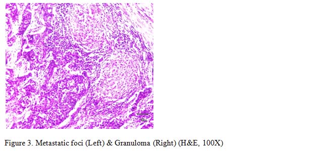
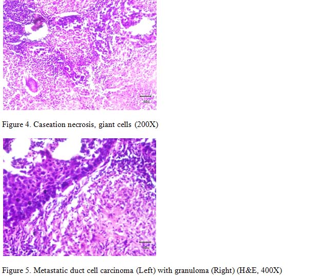
Out of eight nodes three showed tumor metastases and two of the lymph nodes showed epithelioid cell granulomas with Langhans type giant cells and central caseaous necrosis (Figure 3, 4, 5).

Immunohistochemical (IHC) examination revealed overexpression of estrogen & progesterone receptor and negativity for Her-2/neu in the breast lesion. With these findings, diagnosis of “invasive ductal carcinoma, grade II (Nottingham modification of Bloom Richardson grade) with concomitant regional nodal metastasis done and caseating granuloma” found, so “AJCC staging II”was made. Since preoperatively tuberculosis was not suspected and no other clinical feature of TB was found, mantoux test, culture, serology or polymerase chain reaction were not performed. Due to unavailability modified Ziehl–Nielsen staining on axillary tissue and CD68 immunostain for epithelioidhistiocytes not done.

 Discussion

TB remains a major public health problem worldwide. According to the Global Tuberculosis Report 2018, it affected 10 million people in 2017 and is responsible for 1.2–1.4 million deaths globally in HIV-negative patients. It is caused by Mycobacterium tuberculosis and most often affects the lungs. About 23% of the world’s population (1.7 billion people) has latent tuberculosis, with a risk of reactivation of 5–15%.5 Tubercular lymphadenitis is the most common form of extra pulmonary tuberculosis. But isolated axillary tubercular lymphadenitis without any evidence of clinical disease in any other organ is extremely uncommon in adults. Axillary tubercular lymphadenitis can be accounted for by either retrograde spread from the mediastinal nodes or hematogenous spread from a subclinical focus.

An alternative explanation for the co-occurrence of TB and metastasis could be activation of a silent Mycobacterium tuberculosis infection due to immunocompromised state in cancer patients.6 Tubercle bacillus can exist in a state of microbial latency within the macrophage of the granulomas for the lifetime. Factors that disturb host immunity can allow the tubercle bacillus to cause endogenous reinfection.7

In general, TB is diagnosed by clinical history, erythrocyte sedimentation rate, chest X-ray, Ziehl–Nielsen staining, polymerase chain reaction (PCR)based detection of acid fast bacilli (AFB) and others. But a significant proportion of cases of extra-pulmonary TB may be negative for chest X-rays or regular stains for AFB.8

The clinical features of breast carcinoma may reveal nodules and ulcerations,appearance of lymphadenopathies lead us to suspect a metastatic tumor. It is the histological examination that enabled making the differential diagnosis.9 Ina few cases diagnosis have been made through preoperative investigations like cytology or core biopsy. PET-CT as we know is the imaging of choice in certain cancers like lung cancer, its role in breast cancer is limited.8

Breast cancer patientsalso may suffer reactivation of TB during their treatment. It not only disturb the treatment protocol but also the clinical and radiologic findings confuse the follow up process since a malignant and a tubercular lesion may be indistinguishable.7 Axillary lymph node metastasis is the most important factor in the staging of ductal carcinoma and the number of metastatic axillary nodes alter the stage. As tuberculosis also produces nodal enlargement, this can mimic or complicate the staging of malignant disease.6 Therefore, pathologists and lab technicians should also be aware and vigilant in ruling out possible differentials such as metastasis, tuberculosis, fat necrosis, actinomycosis, suture granuloma accordingly.10

 Conclusion

Simultaneous presence of axillary tubercular lymphadenitis and metastatic carcinoma is a significant but rare event that can confuse and complicate the diagnosis, grading, staging and most importantly, the treatment of the disease. It is important for the physician in endemic countries like ours to consider the possibility of a granulomatous disease masquerading as axillary metastasis in order to correctly address both diseases.


 References

  1. Pandey M, Abraham EK, Chandramohan K and Rajan B. Tuberculosis and metastatic carcinoma coexistence in axillary lymph node: A case report. World Journal of Surgical Oncology; 2003;1(3):1-3.
  2. Tulasia NR, Rajub PC, Damodaranb V and Radhikab TS. A spectrum of co-existent tuberculosis and carcinoma in the breast and axillary lymph nodes: Report of five cases.The Breast; 2006;15:437–439.
  3. Caroppo D, Russo D, Merollam F, Ilardi G, Caro MD, Lorenzo PD, Varricchio S, Mascolo M and Staibano S. A rare case of coexistence of metastasis from head and neck squamous cell carcinoma and tuberculosis within a neck lymph node. Diagnostic Pathology; 2015;10(197):1-3.
  4. Kaplan MH, Armstrong D, Rosen P. Tuberculosis complicating neoplastic disease:A review of 201 Cases. Cancer 1974;;33:850-858
  5. Almeida S, Valentim M, Neto C, Cerol M, Boticário M, Santos MI, Gameiro A. Extrapulmonary Tuberculosis Presenting as a Suspected Case of Metastatic Breast Cancer. European Journal of Case Reports in Internal Medicine 2019. DOI:10.12890/2019_001062
  6. Pujani M, Khan S, Hassan MJ, Jetley S and Raina PK. Coexistence of metastatic breast carcinoma and primary tuberculosis in axillary lymph nodes: A report of a rare case; Breast Disease. 2015;35:195–198.
  7. Baslaim MM, Al-Ghamdi MA, Al-Numani TS, Ashour AS and Al-Amoudi SA. Tuberculosis in 7 breast cancer cases: Diagnostic and therapeutic challenges. J Mycobac Dis; 2013;3(3):1-4.
  8. Mukhopadhyay T, Nandi M, Bhattacharya S, and Khan EM. Contralateral contiguous tuberculous lymphadenitis in a case of right breast carcinoma – Diagnostic dilemma. J Can Res Ther.2015;11:102-6.
  9. Ouedraogo AS, Bambara HA, Ademayali FAH, Ramde WN, Sawadogo RJ, Savadogo I, Ouattara S, Barry1 H, Lamien AS, Lompo OM. Anatomo-clinical case: coexistence of tuberculosis with axillary lymph node metastasis in breast carcinoma. Open Journal of Pathology; 2018;8:132-138.
  10. Lee CH. and Sharif SJ. Primary breast tuberculosis (TB) in a patient with known invasive breast carcinoma: A case report. Med J Malaysia.2016;71(3):149-151.

jhc-2019-v-3-n-2-Is-histopathological-kabir-an

Review Article

Is Histopathological  Examination Essential  for the Diagnosis of Psoriasiform Dermatitis?

*Kabir AN,1 Rahman MM 2

  1. *Dr. AKM Nurul Kabir, Associate Professor, Department of Pathology, Bangabandhu Sheikh Mujib Medical University, Dhaka, Bangladesh. kabir56@gmail.com
  2. Mohammad Mosiur Rahman,  Assistant Professor, Department of Pathology, Bangabandhu Sheikh Mujib Medical University, Dhaka, Bangladesh

 *For correspondence

Abstract

Psoriasiform dermatitis encompasses a wide range of diseases, some of which show both clinical and histological overlap. Psoriasis is generally thought to be a genetic disease that is triggered by environmental factors. Psoriasis is characterized by an abnormally excessive and rapid growth of the epidermal layer of the skin.  Abnormal production and an over abundance of skin cells result from the sequence of pathological events in psoriasis, can be shown as a higher Ki-67 index compared with normal appearing, non-lesional skin. Morphometric analysis  of  histological features can also give a quantitative dimension  to histopathology  in diagnosis of psoriasis and in differentiating from other psoriasiform dermatitis. Psoriasis is also regarded as a T-cell mediated disorder, mainly CD4+ (helper/inducer) lymphocytes along with CD8+ subsets are known to occur.  The patient often prove to be a diagnostic dilemma for both dermatologists and pathologists alike. However, clinical features when considered alone may not be reliable, as they vary with both disease duration and treatment.  Though to give a precise diagnosis one relies on clinical correlation, histopathology is essential as it is possible to assign specific diagnosis in most cases with a logical and systematic histopathological approach. Morphometry along with the help of Ki-67 & Cyclin D1 and other immunostains of keratinocytes, and immunophenotyping of T-cell infiltrate, a definitive diagnosis can be made.

 [Journal of Histopathology and Cytopathology, 2019 Jul; 3 (2):151-161]

Key words: Psoriasis, Psoriasiform dermatitis, Morphometry, Ki-67, CD4+, CD8+

 Introduction

Psoriasiform  dermatitis  encompasses a wide range of inflammatory dermatoses, some of which show both clinical and histological overlap.  The term psoriasiform means  that  the lesion either clinically or histopathologically mimic  psoriasis and this group includes: psoriasis- the prototype of psoriasiform dermatitis,1 and others as  seborrheic dermatitis,  pityriasis rubra pilaris (PRP), allergic dermatitis, atopic dermatitis , nummular dermatitis,  lichen simplex chronicus (LSC), prurigo nodularis,  pityriasis rosea (PR), inflammatory linear verrucous  epidermal nevus (ILVEN) and mycosis fungoides (MF). Besides, clinical features in one patient may differ at different times and the diagnosis get obscured.3 As there is varied clinical presentation, a definitive histopathological  diagnosis is essential in the treatment of such inflammatory dermatoses.1 It is a challenging  task even to experienced pathologists to give a precise diagnosis every time and one relies also on clinical correlation. However, a stepwise systematic histopathological approach enables one to reach at a specific diagnosis in most cases.2

The morphometry parameters such as length of rete ridges, the length of dermal papillae and the ratio of length/average width of rete redges are statistically significant in the differentiating  psoriasis from psoriasiform dermatitis and can be used in addition to routine histopathology.3 As the prototype of psoriasiform dermatitis, psoriasis is a hyperproliferative skin disorder with increased epidermal turnover rate and mitotic index, proliferation markers Ki-67 & Cyclin D1 immunostain of keratinocytes can be added as diagnostic tools  to differentiate from other non- psoriasiform dermatitis.4  Expression levels of pRb and p53 were found to be higher in the psoriasis group in a study,  compared with the normal epidermis.5  Psoriasis is an autoimmune skin disease and regarded to be T- cell mediated disorder. CD4+ T-cells are important in initiating and maintaining the pathogenic process of psoriasis but cross-primed CD8+ T-cells are the main effector cells. Mixtures of CD4+ T cells and CD8+ T-cells are found in papillary dermis and epidermis of psoriatic lesion.  Psoriasiform lesions on the other hand show a high proportion of CD4+ T-cells in dermis.  Immunophenotyping of T-cell infiltrate in the lesion can be done in differentiating the cases.6


History of Psoriasis

History of psoriasis begins in Ancient Greece, when psoriasis, leprosy, and other inflammatory skin disorders were believed to be the same condition and the Greeks termed the skin diseases as  psora, lepra and lichen. Psora referred to itch, and lepra from lopos & lepo (the epidermis & to scale respectively). Hippocrates (460-377 BC) used the word lopoi to describe the dry, scaly, disfiguring disorders.  The Old Testament also lumped together many cutaneous   disorders, including leprosy and psoriasis, by the biblical term tsaraat or zaraath. Lepers were considered divinely punished, and cruelty was imposed upon those who were suffered from psoriasis and leprosy alike. Roman medical writer Celsus (25BC-45AD) first described papulosquamous diseases, suggesting as psoriasis one of these. Galen (133-200) first used the term psoriasis, but his description was likely represented seborrheic dermatitis. Gilbert (1797-1866) and Hebra finally distinguished the clinical picture of psoriasis from that of leprosy.7

 Epidemiology

Although psoriasis occurs worldwide, it is a common chronic inflammatory skin disorders affecting 1.5-2% population in the western countries and 1.3% in general population.6 The prevalence of psoriasis is low in certain ethnic groups such as the Japanese.8

Psoriasis can present at any age and has been reported at birth and in older people of advanced age. A bimodal age of onset has been recognized in several large studies. The mean age of onset for the first presentation of psoriasis can range from 15 to 20 years of age, with a second peak occurring at 55–60 years. Henseler and Christophers studied a series of 2147 patients and also reported as two clinical presentations of psoriasis, type I and II. Type 1 begins on or before age 40 years; Type II begins after the age of 40 years. Type I disease accounts for more than 75% of cases. Patients with early onset, tended to have more relatives affected and more severe disease than patients who have type II psoriasis. In addition, strong associations have been reported with human leukocyte antigen (HLA)-Cw6 in patients with early onset of psoriasis. The course and progress of psoriasis is unpredictable. In one study, 39% of patients reported complete remission of disease at the age between one and 54 years. Higher figures have been reported in Japan.8 A family history of the disease is common. Approximately 30% of patients have a first-degree relative with psoriasis, and the risk of psoriasis increases with the number of affected relatives a patient has.9

 Lifestyle and Morbidity

There is a link between cigarette smoking and psoriasis severity. There is sufficient evidence that aggravation of psoriasis is associated with alcohol consumption. Various recent studies report association of diabetes and other cardiovascular diseases to the severe form of psoriasis.10 Recent research suggests that patients with psoriasis have a systemic inflamma­tory state, putting them at increased risk of cardiovascular complications, including metabolic syndrome, peripheral vascular disease, stroke, myocardial infarction, and cardiac death.11  Psoriasis is increasingly being recognized as a disease that not only affects the skin but also has multi-systemic implications. Increasing epidemiological evidence suggests that patients with psoriasis may be more obese compared with the general population. Although the exact mechanism underlying the epidemiological association between psoriasis and obesity is uncertain, researchers have theorized that adipocyte elaboration of pro-inflammatory cytokines may exacerbate psoriasis.12 Many, but not all, studies have shown a positive association between metabolic syndrome and cardiovascular disease (CVD) and psoriasis, especially for (young) patients with moderate to severe disease. A recent study showed that Glyc A, which is a novel biomarker for systemic inflammation  was associated with psoriasis.13  Nearly 60% of psoriasis patients and 40% of psoriatic arthritis patients report their disease as a large problem in their everyday life. Psychosocial limitations of both diseases include enduring low self-esteem, feeling physically unattractive or sexually undesirable and avoiding social activities.14

Etiology

Etiology of psoriasis remains unknowneven though, it is believed to be multifactorial with numerous key components including genetic susceptibility, environmental triggers in combination with skin barrier disruption and immune dysfunction.15 Various data suggest that infection is an important trigger for psoriasis. Streptococcal throat infection can initiate and exacerbate chronic psoriasis.15 Another research has shown that the composition of the cutaneous microbiota is related to many dermatological diseases including  psoriasis, atopic dermatitis, and acne vulgaris.16

Genetic Factors

Epidemiological studies have demonstrated that psoriasis has an important genetic component, where the role of environmental triggers (e.g., stress, mechanical trauma and streptococcal infections) is well documented.  Familial recurrence is also documented and disease concordance is higher in monozygotic than dizygotic twins. So, psoriasis is widely regarded as a multifactorial disorder caused by the interaction between inherited susceptibility alleles and environmental risk factors.17 The molecular genetic basis of psoriasis is complex, however, there are evidence that multiple genes are involved. Seven major psoriasis susceptibility loci have been reported and   a major susceptibility locus for psoriasis is at 6p21, referred to as PSORS1 and is overrepresented in all populations tested.  An association between psoriasis and other loci has also been reported on chromosomes 1p (PSORS7), 1q (PSORS4), 3q (PSORS5), 4q (PSORS3), 17q (PSORS2), and 19p (PSORS6). The strength of associations between such genes and susceptibility to psoriasis, apart from PSORS1, is variable, may relate, in part, to heterogeneity among different populations.8

 Mechanism

Psoriasis is characterized by an abnormally excessive and rapid growth of the epidermal layer of the skin. Abnormal production of skin cells and an overabundance of skin cells result from the sequence of pathological events in psoriasis.  Skin cells are replaced every 7 days in psoriasis rather than the usual about 53 days.18 Exclusive cellular “responsibility” for the induc­tion and maintenance of psoriatic plaques has not been clearly defined. Increased proliferation of keratinocytes and endothelial cells in conjunction with APC/T cell/monocyte/macrophage inflammation leads to the distinct epidermal and vascular hyperplasia that is characteristic of lesional psoriatic skin.15  CD8+ T cells seem to be dominant in the epidermis whereas the CD4 + T cells predominant subset in the dermis.  Activated CD4+ T cells produce a variety of  cytokines  including interleukin-2 (IL-2), TNFα and γINF.  TNFα stimulating keratinocytes may produce  IL -8, a potent T- lymphocyte and neutrophil chemoattractant  and may involved in the formation of Munro microabscess. γINF is believed to play in important role in the initiation of psoriatic lesion.18 In individuals with a genetic predisposition, external stimuli such as trauma (known as Koebner phenomenon), infections, stress, drugs, and alcohol can all trigger an initial episode of psoriasis. This initial trigger activates the innate immune system (Figure 1). Complexes of the antimicrobial peptide LL-37 and host DNA/RNA, both released by keratinocytes after common epidermal damage, activate plasmacytoid dendritic cells (pDCs) to produce large amounts of type I IFNs (α/β).  Subsequently, type I IFNs trigger maturation and differentiation of dermal dendritic cells (dDCs), and these dDCs then stimulate autoreactive T cells. Thereby, psoriatic autoimmune T cells are biased to produce Th1 and Th17/Th22 cells and which induce TNFα,/INFγ and IL-17/IL-22 production respectively. These mediators act on keratinocytes, leading to the activation and proliferation. In addition, type I IFNs directly upregulate IL-22 receptor (IL-22R) on keratinocytes, increasing their responsiveness to IL-22, and inhibits terminal differentiation and induces hyperproliferation of keratinocytes leading to epidermal hyperplasia, resulting psoriasis.19  In psoriasis , along with epidermal changes there is markedly increased dermal vascularity. The dermal vascular changes have been evaluated and shown to be due to change from arterial-type vessels to venules. The vascular changes may also precede the epidermal alterations, as shown in some studies.20

Clinical Features

Psoriasis is a papulosquamous disease with variable morphology, distribution, severity and course. Papulosquamous diseases are characterized by scaling papules and plaques.8  There are five clinical subtypes of psoriasis: vulgaris (plaque), guttate, pustular, inverse, and erythrodermic. The most common variant of psoriasis is plaque psoriasis, which includes approximately 85%–90% of psoriatic patients.15 The lesions are typically distributed symmetrically on the extensor surfaces of elbows, knees, and lumbosacral area and scalp. Psoriasis may also develop at the site of trauma, known as Koebner’s phenomenon.  Guttate psoriasis has drop-shaped lesions.  Inverse psoriasis forms red patches in skin folds. Pustular psoriasis presents as small non-infectious pus-filled blisters. Erythrodermic psoriasis involves widespread inflammation and exfoliation of the skin over most of the body surface. Psoriatic nail disease affects fingernails more commonly than toenails with small pits in the nail plate.8

Clinical Diagnosis

Psoriasis is a clinical diagnosis, and a skin biopsy is usually not necessary for a classic presentation of the disease.9 Diagnosis of psoriasis is usually based on the appearance of the skin. The characteristic lesions are sharply demarcated, scaly, erythematous plaques, papules, or patches on skin that may be pruritic and/or painful. They may be ovoid, round or irregular and often distributed symmetrically.9 Auspitz sign, named after Heinrich Auspitz, where there is pinpoint bleeding on removal of scales from the lesions of psoriasis. The test by which Auspitz sign is elicited by light tangential scratching with the edge of glass slide is called as Grattage test can be used  as clinical tool.21

Microscopic Diagnosis

Histopathology is considered as a “gold standard” for the diagnosis of most dermatological conditions including psoriasis.21 Common histological features in  psoriasis vulgaris are  hyperkeratosis, parakeratosis,  psoriasiform hyperplasia (Figure 2),  Munro microabscess / spongioform pustule of Kogoj,   thin suprapapillary plate, hypogranulosis, dilated capillaries in papillary dermis and dermal inflammation.1  However, other than  Munro microabscess, spongioform pustule of Kogoj and dilated tortuous capillaries in papillary dermis,  the remaining features , such as  parakeratosis,  psoriasiform hyperplasia can be seen  also in chronic eczematous dermatitis, such as atopic dermatitis, nummular dermatitis or allergic contact dermatitis and appear to be “psoriasiform”.18  However, following a systematic approach one can reach at conclusive diagnosis.


Tirumalae described a detail  microscopic description for definitive diagnosis as follows.2

 Scanning magnification

Examination of microscopic sections under scanning magnification forms the first step. The presence of evenly elongated, thin rete ridges with equally long dermal papillae can seen in one condition i.e., psoriasis. All other diseases exhibit an uneven psoriasiform pattern, where rete ridges are of uneven of lengths and thickness with thick supra-papillary plates as in PRP (Figure 3).

 Stratum corneum

The normal basket-weave pattern is lost in most cases and it is common to find parakeratosis. Parakeratosis can be present in small mounds or may be broad and confluent.  Mounds of parakeratosis can be present throughout the epidermis in conditions like psoriasis and dermatophytosis, in seborrheic dermatitis confined to the infundibular ostia or seen in both locations in PRP.  The classic “checkerboard” pattern i.e., parakeratosis staggered in both vertical and horizontal fronts is seen in PRP. Confluent parakeratosis is seen in dermatophytosis, syphilis, pityriasis lichenoides chronica (PLC) and most importantly, in MF. A sharply defined thick zone of parakeratosis alternating with orthokeratosis and mild papillomatosis are features of ILVEN.

Epidermis

Diminished or absent granular layer is seen in classic psoriasis, but is not a constant feature. Significant amounts of plasma in the scale crust goes against a diagnosis of psoriasis, and  large amounts of plasma and a rapidly proliferating epidermis are hostile to fungi. The finding of spongiotic vesicles in a psoriasiform dermatitis are seen in allergic contact/nummular dermatitis and seborrheic dermatitis. Spongiotic psoriasiform pattern is also encountered in patch-stage of MF.

 Changes in Papillary Dermis

Psoriasis is characterized by thin supra-papillary plates and elongated dermal papillae with dilated, tortuous capillaries. The presence of extravasated erythrocytes is a sign of acuteness and is seen in eruptive psoriasis and PR. Vertical streaks of collagen with uneven psoriasiform acanthosis and compact orthokeratosis is diagnostic of LSC. However, changes of LSC can be superimposed on any chronic, itchy dermatitis including psoriasis. In contrast, thin haphazard, wiry bundles of collagen are seen in the papillary dermis in PLC and MF.

 Nature of Infiltrate

Most of the psoriasiform dermatoses are dominated by lymphocytes in the dermis. When eosinophils are present among them, it indicates allergic/contact dermatitis or a drug eruption.  However, eosinophils are not a pre-requisite for diagnosis of either of these conditions. Histiocytes and plasma cells in a psoriasiform dermatitis with scale crusts give hints for secondary syphilis. Superficial and deep infiltrates are seen in syphilis and lichen striatus (LS).  In LS, there is a psoriasiform lichenoid pattern with deep infiltrates of lymphocytes along the adnexae, especially around the eccrine glands.

 Morphometry

Morphometric analysis of histological features can give a quantitative dimension to histopathology. The results of morphometry parameters such as length of rete pegs, the length of dermal papillae and the ratio of length/average width of rete pegs  can help in achieving the accurate diagnoses of psoriasis and psoriasiform dermatitis. In one study using eye-piece micrometer with the light microscope without special software and found statistically significant results in differentiating psoriasis from psoriasiform dermatitis. The mean length of rete pegs was 1.74 times greater in psoriasis when compared to psoriasiform dermatitis. The ratio of average length and width of rete pegs was considered representative of shape of rete pegs. The higher ratio of length over average width of rete pegs confirms the histopathological finding of slender long rete pegs, which favors the diagnosis of psoriasis.

The dermal vascular changes have been evaluated in different studies by electron microscopy and shown to be due to change from arterial-type vessels to venules. The vascular changes may also precede the epidermal alterations, as shown in some studies. Morphometric parameters of papillary dermal vessels in psoriasis and to compare these with psoriasiform lesions can also be used. Morphometric evaluation of dermal vasculature in psoriasis has shown increased endothelial and luminal volume of vessels compared to control subjects. On light microscopic evaluation of CD34 immunostained sections, biopsies from psoriasis showed much greater microvascular staining in the papillary dermis in comparison to psoriasiform dermatitis.20

 Role of Immunohistochemistry

  Ki-67 and Cyclin D1 Immunostaining

As Psoriasis is a hyperproliferative skin disorder with increased epidermal turnover rate and mitotic index,  proliferation markers Ki-67 (Figure 4) and Cyclin D1 immunostaining  can be used to differentiate psoriasis from non-psoraisis psoriasiform dermatitis( NPPD ) (Figure 5). In a study Ki-67 and Cyclin D1 positive cells were counted per mm2 of suprabasal and total epidermal area by Sezer. et al. 4

Psoriatic lesions have been shown to reveal a higher Ki-67 index compared with normal appearing, non-lesional skin.  In psoriasis group, suprabasal Ki-67 positive cells were more than 75% of the total epidermal Ki-67 positive cells in the epidermis (including basal and suprabasal cell population) in psoriasis group, whereas this ratio was lower than 75% (cut-off value) in NPPD group. The suprabasal/total epidermal count ratio for Cyclin D1 immunostaining was higher in the psoriasis group compared with the NPPD group, but a cut-off value to distinguish between these two was not detected, suggesting that Ki-67 is a more sensitive marker than Cyclin D1 in terms of the presence of a cut-off value and could be a useful tool for dermatopathologists to differentiate psoriasis from other psoriasiform dermatitis.4

 pRb and p53 Immunostaining

Expression levels of pRb and p53 were found to be higher in the psoriasis group  in a study,  compared with the normal epidermis.  The phosphorylation of pRb is associated with cyclin D1, cyclin E and P16.  pRb phosphorylation in psoriasis is incresed by the activity of cyclin D1 and Cyclin E and showed a higher expression of pRb than that of normal epidermal expressions.  Total basal layer cell counts for p53 expression were found to be significantly higher in psorisis group compared with the normal group.5 It is postulated that increased expression of p53 in psoriatic skin is a physiological reaction indicating the attempt  to counteract the proliferation and to repair DNA errors, and is most often expressed as an increased number of mitoses.22

 CD4+ and CD8+ cell distribution

Psoriasis also known as a skin disorder with immunological factors playing an important role in its pathogenesis, and lymphocytic infiltrates, mainly CD4+ and CD8+ cells provide a major contribution in the initiation and maintenance of the lesion. Psoriasis is an autoimmune skin disease characterized by T-cell mediated hyperproliferation of keratinocytes. It is regarded as a T-cell mediated disorder, mainly CD4+ (helper/inducer) lymphocytes along with CD8+ (suppressor/cytotoxic) subsets are known to be associated. T-lymphocytes in psoriatic lesion are in an activated state with expression of HLA-DR and IL-2 receptor. Mixtures of CD4+ T-cells and CD 8+ T-cells are present in papillary dermis and epidermis of psoriatic lesion. T-cell also plays a role in the pathogenesis of psoriasiform lesions, and reported a high proportion of CD4+ T-cells in the dermis on immunohistochemical analysis.6

A comparative study was done by Rana. et al to see the infiltration of CD4+ and CD8+  cells in psoriasis and psoriasiform lesions. The inflammation in the epidermis was present in about half of psoriasis cases and predominant cells were polymorphs. In the upper dermis, inflammation was evident in all cases with predominant cells were lymphocytes. Inflammation in the lower (reticular) dermis was present with mostly lymphocytes.

In psoriasiform lesions the inflammation was present in the epidermis in only few cases showing mixtures of polymorphs and lymphocytes. Within the upper dermis, all cases showed inflammation with predominant of lymphocytes. Few cases showed inflammation in the lower dermis with lymphocytes as majority of the cells. Immunohistochemical staining was done to see the distribution of CD4+ and CD8+ cells.  the distribution of CD4 positive cells was seen in psoriasis patients.  In the epidermis of psoriasis cases only one fifth were positive for CD4+ cells. In the upper dermis, CD4+ cells were present in 92% cases.  In the lower dermis about half of cases were positive for CD4+ cells.

D 8+ positive cells on the other hand were seen in the epidermis in 40% cases.  In the upper dermis, all the cases showed CD8+ positive cells.  In the lower dermis, CD8+ positive cells were found in large number of cases.

An immunophenotyping of the inflammation of psoriasiform lesions showing, CD4+ cells as predominant cells in the upper dermis. In lower dermis area showed 16% cases having CD4+ cells.

Various studies have shown that CD4+ cells are less frequent in psoriatic epidermis. In epidermis CD8+ cells were seen in 40% cases, and 8% in psoriasiform lesions. The difference was statistically significant.  In the upper dermis, one study has shown the mixtures of CD4+ and CD8+ T-cells in psoriasis, whereas psoriasiform group showed that CD4+ cells in abundance.

The difference in amount and pattern of CD4+ and CD8+ T-cells in the various compartments is helpful in differentiating between psoriasis and psoriasiform lesions especially in cases with borderline morphology.6

 Conclusion

Psoriasis, the prototype of psoriasiform dermatitis, is a genetically determined, inflammatory, and proliferative disease of the skin characterized by dull red, sharply demarcated scaly plaques. The two clinical signs, Auspitzs sign and the Grattage test have been described as pathognomonic of psoriasis when biopsy is not done.  However, these signs are present only in untreated patients. Psoriasis has many different clinical differentials as other psoriasiform lesions. Besides, the same patient can present at different times with a different clinical presentation.  Since satisfactory management of the condition requires both symptomatic and specific therapy, it is essential to reach a definitive diagnosis. The recurrent nature and prognosis of psoriasis differs from that of psoriasiform dermatitis, thus, further highlighting the importance of reaching the correct diagnosis. Clinical features, considered alone, may not be reliable. Histopathology is regarded as a gold standard for the diagnosis of most dermatological conditions including psoriasis and constitutes definite hard evidence, which can be preserved for future review.  Though to give a precise diagnosis one relies on clinical correlation, histopathology is essential as it is possible to assign specific diagnosis in most cases with a logical and systematic histopathological approach.  In selected cases morphometry, Ki-67 & Cyklin D1, pRb and p53 immunostaining of keratinocytes can be added when needed. The pattern of T-cell infiltrate by immunophenotyping can give an additional diagnostic clue.

Acknowledgement

The authors are grateful to Faisal Kabir Taj for his help in preparing the figure 1.

 References

  1. Jayalakshmy PL, Babitha AM, Sankar S, Nandakumar G. Histopathological spectrum of Psoriasiform dermatitis. Journal of Pathology of Nepal 2016; 6(12): 975 -980.
  2. Tirumalae R . PsoriasiformDermatoses: Microscopic Approach,Indian J Dermatol. 2013 58(4): 290–293. doi: 4103/0019-5154.113945.
  3. Shirish S. Chanadanwale, Narayanan K. Panicker, Sushmah P. Kulkarni, Komal R. Shah, Kumar H, Yugal K. Sharma1, Sukanya Pal, Morphometry analysis of psoriasis and psoriasiform dermatitis: A retrospective study of 50 cases.  Medical Journal of  D.Y. Patil University 2015; 8(1): 43-47.
  4. Sezer E, Böer-Auer A, Cetin E, Tokat F, Durmaz E, Sahin S and Ince U. Diagnostic utility of Ki-67 and Cyclin D1 immunostaining in differentiation of psoriasis vs. other psoriasiform dermatitis. Dermatol Pract Concept. 2015;5(3): 7-13. 2015;5(3):2ul; 5(3):doi: 5826/dpc.0503a02.
  5. Kim SA, Ryu Y W, Kown J I, Choe M S, Jung J W, Cho J W. Differential expression of cyclin D1, Ki‑67, pRb, and p53 in psoriatic skin lesions and normal skin. MolecularMedicine  Reports 2018;17: 735-742,   DOI: 10.3892/mmr.2017.8015.
  6. Rana S, Zeeba J S, Sujata J, Madhur K: A comparative study of psoriasis and psoriasiform lesion on basis of CD4 and CD8 cell infiltration. Our Dermatol Online. 2012; 3(4): 292-297.
  7. Cowden A., Van Voorhees A.S. (2008) Introduction: History of psoriasis and psoriasis therapy. In: Weinberg J.M. (eds) Treatment of Psoriasis. Milestones in Drug Therapy. Birkhäuser Basel, pp 1-2. DOIhttps: doi.org/10.1007/978-3-7643-7724-3_1.
  8. Langley R G B, Krueger G G, Griffiths C E M, Psoriasis: Epidemiology, Clinical Features, and Quality of Life. Ann Rheum Dis. 2005;64(Suppl II):ii18 23.doi: 10.1136/ard.2004.033217.
  9. Grace W. Kimmel and Mark Lebwohl. Psoriasis: Overview and Diagnosis.Bhutani T et al. (eds.), Evidence-Based Psoriasis, Upsoriasiformdermatitisates in Clinical Dermatology, https://doi.org/10.1007/978-3-319-90107-7_1.
  10. Khan ZA, Malik LM,  Jahangir M,  Prevalence of smoking, alcohol, and comorbid conditions in psoriasis. Journal of Pakistan Association of Dermatologists 2010; 20:212-216.
  11. Sako E, Famenini S, Jashin J Wu.  Trends in Type of Original Psoriasis Publications by Decade, 1960 to 2010.   Perm J 2016 Fall;20(4):15-060. http://dx.doi.org/10.7812/TPP/15-060.
  12. Armstrong AW, Harskamp CT, Armstrong EJ. The association between psoriasis and obesity: a systematic review and meta-analysis of observational studies. Nutrition and Diabetes (2012) 2, e54; doi:10.1038/nutd.2012.26..
  13. Samuel T. Hwang, Nijsten T and Elder J T. Recent Highlights in Psoriasis ResearchJournal of Investigative Dermatology 2017; 137(3):550 -556; doi:10.1016/j.jid.2016.11.007.
  14. Busse K, Liao W.Which Psoriasis Patients Develop Psoriatic Arthritis? Psoriasis Forum. 2010; 16(4): 17–25.
  15. Ayala-Fontánez N,  Soler D C, McCormick TS.   Current Knowledge on Psoriasis and Autoimmune Diseases.    Psoriasis: Targets and Therapy 2016;6: 7–32;.org/10.2147/PTT.S64950.
  16. Benhadou F, Mintoff D, Schnebert B, Thio H B. Psoriasis and Microbiota: A Systematic Review. Diseases 2018, 6, 47; doi:10.3390/diseases6020047.
  17. Capon F. The Genetic Basis of Psoriasis. Int J Mol Sci. 2017;18(12):2526. Published 2017 Nov 25. doi:10.3390/ijms18122526.
  18. Mobini N, Toussaint S, Kamino H: Noninfectious Erythematous, Papular, and Squamous diseases. In Lever’s Histopathology of the Skin, 10 th Edition. Edited by Elder DE, Elenitsas R, Johnson BL, Murphy GF, Xu X.  India, Woltrs Kluwer, 2009, pp 169-203.
  19. Flatz L, Conrad C. T-cell-mediated inflammation in psoriasis: pathogenesis and targeted therapy.Psoriasis: Targets and Therapy 2013; 3:1-10. org/10.2147/PTT.S26339.
  20. GuptaS,  KaurM,  Gupta R, SinghS, PantL, SinghP P.Dermal Vasculture in Psoriasis and Psoriasiform dermatitis: a  Morphometric study. Indian J Dermatol. 2011 Nov-Dec; 56(6): 647–649. doi: 10.4103/0019-5154.91820: 10.4103/0019-5154.91820
  21. Mehta S, Singal A, Bhattacharya SN. A  study of clinicohistopathological correlation in patients of  psoriasis and psoriasiform dermatitis. Indian J Dermatol Venereol Leprol 2009;75:100.
  22. Baran W, Szepietowski J. C, Szybejko-Machaj G. Expression of p53 protein in psoriasis. Acta Dermatoven APA 2005;14(3):79-83.