Imprint Cytology in the Diagnosis of Upper Gastrointestinal Lesions

Imprint Cytology in the Diagnosis of  Upper Gastrointestinal Lesions

*Keya  SA,1 Saha NK,2 Alam MJ,3 Ullah P,4 Shariar S,5 Hira AD,6 Ahmed NU,7 Islam N8

 Abstract

Endoscopic biopsy and cytology are well established techniques for the diagnosis of upper gastrointestinal tract malignances. Imprint cytology is a touch smear preparation which helps to diagnose malignant cell easily. This cross sectional study was done to see imprint cytological patterns of benign and malignant lesions of upper gastrointestinal tract,  to find out histopathological diagnoses and to compare imprint cytological diagnoses with histopathological diagnoses. Biopsy materials from 100 patients were obtained by endoscopist using video endoscope and were subjected to imprint smear technique followed by histopathological examination of the same materials. Cytological and histopathological diagnoses were compared using appropriate statistical methods. Among the 100 cases, 57(57%) were male & 43(43%) were female with a mean age of 56.7 years.  Out of 100 cases, gastric, esophageal and duodenal lesions were 63(63%), 32(32%) and 5(5%) respectively. Misinterpretation on cytological evaluation was seen in only 6 cases. The overall sensitivity, specificity, positive predictive value, negative predictive value and diagnostic accuracy of imprint cytology were 98.46%, 91.42%, 95.52%, 96.97% and 96% respectively. The findings of the present study indicate that imprint cytological method is a reliable technique for rapid diagnosis of upper GIT lesions as it is simple, accurate and cost effective. Diagnosis can be provided within an hour of endoscopic procedure by the imprint smear technique.

[Journal of Histopathology and Cytopathology, 2018 Jan; 2 (1):23-29]

 Keywords:  Imprint  cytology,  Upper  Gastrointestinal Lesions


  1. *Dr. Shamim Ara Keya, Lecturer of Pathology, Shaheed Suhrawardy Medical College, Dhaka. keyaparash@gmail.com
  2. Professor Dr. Naba Kumar Saha, Professor and Head, Department of Pathology, Sylhet MAG Osmani Medical College, Sylhet.
  3. Md. Jahangir Alam, Associate Professor and Head, Department of Gastroenterology, Sylhet MAG Osmani Medical College, Sylhet.
  4. Parash Ullah, Medical Officer, Department of Medicine, Shaheed Suhrawardy Medical College Hospital, Dhaka.
  5. Sakib Shariar, Pathologist, Bangladesh Railway Hospital, Chittagong.
  6. Ananda Dyuti Hira, Pathologist, Khulna Medical College, Khulna.
  7. Nasir Uddin Ahmed, Pathologist, Faridpur Medical College, Faridpur.
  8. Nazmul Islam, Assistant Professor of Pathology, Army Medical College, Comilla.

*For correspondence

Introduction

Any portion of the gastrointestinal tract may be affected by malignancy. The highest incidence of malignancy is in the esophagus, stomach and colorectal region. In fact esophagogastric and colorectal malignancies are amongst the commonest cancers in humans.1 Worldwide, gastric adenocarcinoma is the second most common cancer.2 Endoscopic biopsy and cytology are well established techniques for the diagnosis of upper gastrointestinal tract malignancies. Now a days, various cytologic techniques like brush cytology, crush preparation, touch smear or imprint cytology are commonly used along with routine endoscopic biopsy.3

Imprint cytology from endoscopic biopsy is highly sensitive and specific technique for the diagnosis of upper GI malignancy. Imprint cytology showed an overall accuracy of 97.6%, 100% and 100% for the diagnosis of malignancies of esophagus, stomach and duodenum respectively.4  But there is no systemic study on imprint cytology of gastrointestinal lesions in our country. Imprint cytology can act as an adjunct to histopathology as it increases the diagnostic efficacy and saves time but definitely it cannot replace histopathology as chances of false positives are high.5  Imprint cytology is a special variation of applied cytology that can be used for various purposes.6 Sometimes endoscopic biopsy fails to make a definitive histological diagnosis due to inadequate tissue from small lesion where tissues are distorted during histological processing; in that case imprint cytology may be useful in diagnosing upper gastrointestinal tract malignancy.7 In this study, imprint smears were taken before keeping the endoscopic specimens in fixative for histopathological examination. Endoscopic biopsy is an essential part of the evaluation of gastrointestinal pathology. In the diagnosis of upper GI lesions, histopathological examination is considered gold standard but is time-consuming  when compared to cytology. Most gastroenterologists and patients would like to get an immediate opinion regarding the adequacy of biopsy and nature of the lesion. Imprint cytology is a simple, rapid, reliable and cost effective method and it alone can serve as a useful tool for an immediate diagnosis within an hour after endoscopic procedure. Imprint cytology lets the surgeon to plan a therapeutic strategy approximately one week earlier.4,8  With this background, this study was designed to find out imprint cytological patterns of benign and malignant lesions of upper gastrointestinal tract, to find out histopathological diagnoses and to compare imprint cytological diagnoses with histopathological diagnoses.

 Methods

This study was a cross sectional study and carried out on 100 patients in the department of Pathology, Sylhet MAG Osmani Medical College, Sylhet in collaboration with the department of Gastroenterology, Sylhet MAG Osmani Medical College Hospital, Sylhet, Bangladesh from 1st January 2015 to 31st December 2015. During endoscopic examination by using video endoscope, biopsy was taken. Imprint smears were prepared from all biopsy samples. Imprint smear was done by placing tissue on the slides from the biopsy forcep with the help of a fine needle.  After placing tissue on the slides, thin smears were prepared by gently rotating the tissue with the needle. Then smears were fixed in 95% ethyl alcohol and stained with Papanicolaou stain.The same tissue after smear preparation was collected for histopathological examination. The tissue was examined thoroughly in day light  with particular emphasis on number, colour, consistency, presence or absence of hemorrhage and necrosis. Tissue was embedded as such and was placed in bottle containing 10% formalin with proper tagging and was kept for overnight fixation.  Routine tissue processing with paraffin impregnation was done and sections were stained with haematoxylin and eosin. The diagnosis of upper gastrointestinal tract malignancy was done according to the cytopathological and histopathological findings and grading was done by WHO classification. Then imprint smear diagnosis was compared with the histopathological diagnosis. The efficacy of imprint cytology was determined by sensitivity, specificity, positive predictive value, negative predictive value and accuracy.

 Results

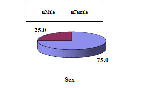
Among the 100 patients, the age ranged from 21 to 100 years with a mean of 56.7 years. In this study, 57(57%) were male and 43(43%) were female with male to female ratio of 1.33:1. The highest number of patients  30(30%) was in the age group 61-70 years followed by 27(27%) in 41-50 years age group & 21(21%) in 51-60 years age group. In our study, 63% (63 cases) were located in the stomach, 32 % (32 cases) were in the esophagus, and 5% (5 cases) were in the duodenum. The most common presenting symptoms were pain and vomiting in case of gastric lesions and dysphagia in case of esophageal lesions.


Table I:  Imprint cytology and corresponding histopathological diagnosis in contingency (cross) table

 Cytological diagnosis


No


Histopathological diagnosis

T


o


t


a


l

Adenocarcinoma Squamous cell carcinoma Chronic non atrophic gastritis Suggestive of adenocarcinoma Chronic gastric ulcer Chronic duodenitis Dysplasia Suggestive of squamous cell carcinoma Chronic esophagitis Hyperplastic polyp Gastric adenoma
Adenocarcinoma 35 34 1 35
Squamous cell carcinoma 25 25 25
Chronic  gastritis 24 23 1 24
Suspicious of adenocarcinoma 5 4 1 5
Chronic gastric ulcer 3 3 3
Suspicious of squamous cell carcinoma 2 1 1 2
Chronic duodenitis 2 2 2
Normal findings 2 1 1 2
Chronic esophagitis 1 1 1
Dysplasia 1 1 1
Total 100 35 25 24 4 4 2 2 1 1 1 1 100


Ulcerative (41%) and ulceroproliferative lesions (41%) were the most common endoscopic findings.  Proliferative lesion was found in 17% of cases and benign polyp was noted in 1% of cases. In the present study, the most common lesion diagnosed by imprint cytology was adenocarcinoma (35%) followed by squamous cell carcinoma (25%) and chronic gastritis (24%) (Table-1). Histopathological evaluation revealed that the most common tumor was adenocarcinoma (35%) followed by squamous cell carcinoma (25%), chronic non atrophic gastritis (24%), suggestive of adenocarcinoma (4%), chronic gastric ulcer (4%), chronic duodenitis (2%),  dysplasia (2%) & suggestive of squamous cell carcinoma, chronic esophagitis, hyperplastic polyp, gastric adenoma  1 case each  (Table I).

Complete correlation between imprint cytological and histopathological diagnosis was obtained in 94(94%) cases (Table II) and 6 cases were misinterpreted on cytological examination. Out of six misinterpreted cases, two cases were diagnosed as normal findings on imprint smears which were subsequently diagnosed as adenocarcinoma in one case and chronic gastric ulcer in another case on histopathology. One case was diagnosed as adenocarcinoma on imprint smears which was subsequently diagnosed as chronic non atrophic gastritis on histopathology. Another one case was diagnosed cytologically as chronic gastritis was diagnosed histologically as hyperplastic polyp. Cytological diagnosis of suspicious of squamous cell carcinoma in one case was confirmed as moderate dysplasia by histopathology. Cytologically one case was interpreted as suspicious of adenocarcinoma which was diagnosed as gastric adenoma on histopathological examination.


Table II:  Correlation of histopathological and cytopathological diagnoses


Histopathological diagnosis No. of Cases Cytological correct diagnosis Percentage
Adenocarcinoma 35 34 97.05
Squamous cell carcinoma 25 25 100
Chronic nonatrophic gastritis 24 23 95.83
Suggestive of adenocarcinoma 4 4 100
Chronic gastric ulcer 4 3 75
Chronic duodenitis 2 2 100
Dysplasia 2 1 50
Suggestive of Squamous cell carcinoma 1 1 100
Chronic esophagitis 1 1 100
Hyperplastic polyp 1 0 0
Gastric adenoma 1 0 0
Total 100 94 94

Statistical evaluation of imprint cytology was done (Table III) and validity of imprint cytology like sensitivity, specificity, PPV, NPV and accuracy were found as 98.46%, 91.42%, 95.52%, 96.97% and 96% respectively (Table IV).

Table III:  Statistical evaluation of cytopathological diagnosis of 100 cases of  upper GIT lesions


Histopathological diagnosis                               Cytopathological Diagnosis
Disease positive (Malignant) Disease negative(Benign)
Positive(Malignant)


65


TP 64 FP 3
Negative(Benign)


35


FN 1 TN 32
Total 100 Total 65 Total 35


TP= True positive, TN= True negative, FP= False positive, FN= False negative.


Table IV:  Validity of cytopathological diagnosis of 100 cases of upper GIT lesions.


Sensitivity Specificity PPV NPV Accuracy
98.46% 91.42% 95.52% 96.97% 96%


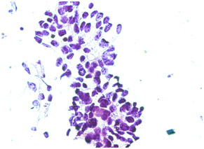
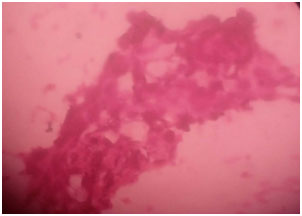
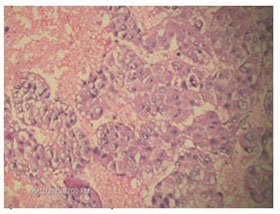
Figure 1. Photomicrograph of imprint cytology of adenocarcinoma (Papanicolaou stain, x40).


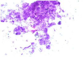
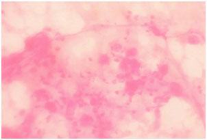
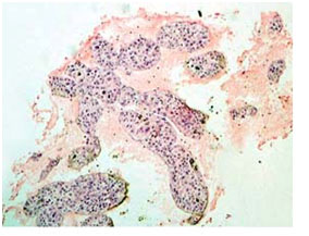
Figure 2. Photomicrograph of imprint cytology of squamous cell carcinoma (Papanicolaou stain, x40).


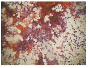
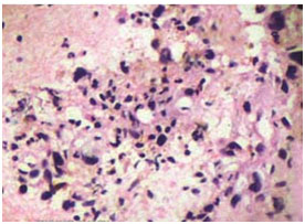
Figure 3.  Photomicrograph imprint cytology of chronic gastritis (Papanicolaou’s stain, x40).


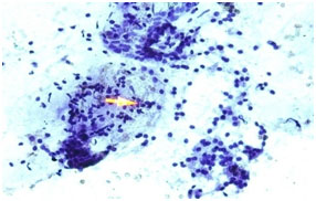
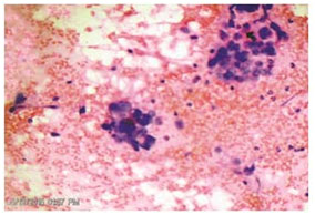
Figure 4. Photomicrograph of imprint cytology of gastric ulcer (Papanicolaou stain, x40).

Discussion

The primary role of gastrointestinal tract cytology is the detection of cancer. Gastrointestinal cytology is still in its early stages of development and few studies have described the role of brush cytology and imprint cytology in interpretation of upper GI biopsy.

The age of study patients ranged from 21 years to 100 years with a mean of 56.7 years.  Of the 100 patients, 57(57%) were male and 43(43%) were female with male to female ratio of 1.33:1.  This observation is similar to other studies who also observed that the frequency of gastrointestinal lesions was more in male.4,9  Highest number (30%) of gastrointestinal lesions were found in the age group 61 to 70 years followed by 27(27%) in 41 to 50 years age group and 21(21%) in the age group of 51 to 60 years which have consistence with the study of Vijayanarasimha D et al9  who observed highest number in fifth to seventh decades of life with a mean age of 55 years.  In the present study, 63% lesions were in the stomach, 32% were in the esophagus and 5% were in the duodenum which are also close to the findings of the  study  done by Vijayanarasimha D et al9  who observed that out of 110 cases, 52(47.3%) were gastric, 45(40.9%) were esophageal and 13(11.8%) were duodenal lesions. The clinical presentations of study patients were upper abdominal or epigastric pain, dysphagia, vomiting, anorexia, distended abdomen and sometimes abdominal mass.  Abdominal or epigastric pain was the commonest symptom in case of gastric and duodenal lesions and dysphagia in case of esophageal lesions. This clinical observation is similar to the clinical findings of the study done by Vijayanarasimha D et al.9 In our study, endoscopic findings of upper GIT lesion were ulcerative (41%), ulcero-proliferative (41%), proliferative (17%) and polypoid (1%).  In this study, among 100 cases of upper GIT lesions, 65(65%) were malignant and 35(35%) were benign. Diagnostic accuracy of imprint cytology of esophageal, gastric and duodenal lesions were 96.88%, 95.24% and 100% respectively which corresponds to or even better than some of the other studies.4,5,9,10,11 Histopathological evaluation of biopsies from 100 cases revealed that the most common lesion  was adenocarcinoma (35%) followed by squamous cell carcinoma(25%), chronic  non atrophic gastritis (24%), suggestive of adenocarcinoma(4%), chronic gastric ulcer (4%), chronic duodenitis (2%) and dysplasia (2%). Chronic esophagitis, hyperplastic polyp, gastric adenoma constituted  one case each.

Complete correlation between imprint cytological and histopathological diagnosis was obtained in 94(94%) cases and 6 cases were misinterpreted on cytological examination. One false negative case found in this study that was observed in gastric lesion. This false negative result may be due to subepithelial location of the lesion which was missed on imprint smear.  False negative diagnosis in imprint cytology was also observed in subepithelial lesions studied by Asha M et al.10 On endoscopic examination, ulceroproliferative lesion was seen in two cases in the gastric antrum. In one case, imprint cytology smears showed anaplastic epithelial cells and a diagnosis of adenocarcinoma was made. Histopathological examination showed chronic non atrophic gastritis and did not reveal any evidence of malignancy. Other case was interpreted as suspicious of adenocarcinoma by imprint smear which was subsequently diagnosed as gastric adenoma with dysplastic change by histopathology. On endoscopic examination, an ulceroproliferative lesion was seen at the lower end of esophagus which was interpreted as suspicious of squamous cell carcinoma on imprint smear but histopathological examination revealed moderate dysplasia. In the present study, three false positive diagnoses were made by imprint cytology. False positive diagnosis was also reported by other researchers and they concluded that the false positive diagnosis was due to regenerative atypia.9,11

The highest correlation (100%) was observed between imprint cytology and histopathology in the diagnosis of squamous cell carcinoma, suggestive of adenocarcinoma, suggestive of squamous cell carcinoma, chronic duodenitis and chronic esophagitis.

The overall sensitivity, specificity, positive predictive value, negative predictive value and diagnostic accuracy of imprint cytology in the diagnosis of upper gastrointestinal lesions were 98.46%, 91.42%, 95.52%, 96.97% and 96% respectively which are similar to other studies.4,9

 Conclusion

The findings of the present study indicate that imprint cytology is a reliable technique for rapid diagnosis of upper GIT lesions as it is simple, accurate and cost effective. Diagnosis can be provided within an hour of endoscopic procedure by the imprint smear technique. So, gastroenterologist or surgeon can take a therapeutic decision approximately one week earlier.

References

  1. Conrad R, Castelino-Prabhu S, Cobb C, Raza A. Role of cytopathology in the diagnosis and management of gastrointestinal tract cancers. Journal of Gastrointestinal Oncology 2012; 3: 285-98.
  2. Zhang XF, Huang CM, Lu HS et al. Surgical treatment and prognosis of gastric cancer in 2613 patients. World Journal of Gastroenterology 2004; 10: 3405-08.
  3. Batra M, Handa U, Mohan H, Sachdev A. A Comparison of cytohistologic techniques in diagnosis of gastroesophageal malignancy. Acta Cytologica 2008; 52: 77-82.
  4. Sanjeevreddy M, Kittur SK, Jakareddy RB, Patil SY, Yelikar BR. Role of imprint cytology in the diagnosis of upper gastrointestinal tract lesions. Indian Journal of Public Health Research and Development 2013; 4: 190-95.
  5. Sharma P, Misra V, Singh PV, Misra SP, Gupta SC. A correlative study of histology and imprint cytology in the diagnosis of gastrointestinal tract malignancies. Indian Journal of Pathology & Microbiology 1997;40:139-146.
  6. Muller HA. Imprint cytology, advantages and possibilities. Entrez Pub Med 1976; 94: 7-10. [Online]
  7. Lal N, Bhasin DK, Malik AK, Gupta NM, Singh K, Mehta SK. Optimal number of biopsy specimens in the diagnosis of ca esophagus. Gut 1992; 33: 724-26.
  8. Cubukçu A, Gönüllü NN, Kaçar SO, Alponat A, Paksoy N. Imprint cytology in the endoscopic diagnosis of gastrointestinal malignancies. Hepatogastroenterology 2002; 49: 198-200.
  9. Vijayanarasimha D, Mahadevappa A, Manjunath GV, Sunila R. Imprint cytology: A diagnostic aid in interpretation of upper gastrointestinal endoscopic biopsies. Journal of Digestive Endoscopy 2014; 5: 144-148.
  10. Asha M, Divya V, Manjunath GV, Sunila R. Application of imprint cytology in interpretation of esophageal biopsies. Journal of Evolution of Medical and Dental Sciences 2013; 2: 4350-7.
  11. Mysorekar VV, Dandekar CP, Satyaprakash BS, Sarkar A. Role of imprint cytology in the diagnosis of gastrointestinal tract malignancies. Indian Journal of Pathology & Microbiology 2003; 46: 37- 43.


Computed Tomogram Guided Fine-Needle Aspiration Cytology of Lung and Mediastinal Masses: A Study of 166 Cases

Computed Tomogram Guided Fine-Needle Aspiration Cytology of Lung and Mediastinal Masses: A Study of 166 Cases

*Alam MA,1 Islam MR,2 Haque MR, Nath SK4

 Abstract
Computed tomogram guided fine needle aspiration cytology (FNAC) is an important and useful investigation to differentiate between benign and malignant lesions of lung and mediastinum. To evaluate the lung and mediastinal masses and to analyze and compare the results with cytological findings, 166 patients were retrospectively studied who underwent CT guided FNAC over a period of January 2015 to December 2016. The study was carried out in patients who presented with respiratory symptoms with a localized lung and mediastinal masses which were confirmed by radiologically was sent for FNAC. 155 cases of lung masses and 11 cases of mediastinal mass were included in this study. Patients’ age ranged from 15 to 95 year and the male to female ratio was 4:1. Radiologically, out of 166 cases, 140 cases were diagnosed as malignant, 8 cases as benign and 18 cases as inflammatory lesions. Cytologically, 146 cases were diagnosed as malignant, 20 cases were benign inflammatory lesion. Most common lung malignancy was squamous cell carcinoma (72 cases) followed by adenocarcinoma (32 cases), small cell carcinoma (10 cases), large cell carcinoma (8 cases), 18 cases of lung metastasis were seen. Compared to biopsy, CT guided FNAC shortens the diagnostic interval and helps in differentiating lung malignancy into different cytopathological types which aids in proper management of the malignant lesion. Out of 11 mediastinal masses 6 cases were malignant lymphoma, 3 cases specific inflammatory lesions (tuberculosis) and 2 cases was non-specific inflammatory lesions.

[Journal of Histopathology and Cytopathology, 2018 Jan; 2 (1):19-22]

Keywords: Computed tomogram, Cytology, Guided FNAC, Lung mass, Mediastinal mass.


  1. *Dr. Md. Ashraful Alam, Associate Professor, Department of Pathology, Rangpur Medical College. drashraful09@gmail.com
  2. Md. Rezaul Islam, Senior Consultant, Radiology & Imazing, Sadar Hospital, Nilphamari.
  3. Md. Rashedul Haque, Associate Professor, Department of Biochemistry, Rangpur Medical College.
  4. Professor Swapan Kumar Nath, Department of Radiotherapy, Rangpur Medical College.


* For correspondence

 Introduction

A Computed tomography (CT) guided fine needle aspiration cytology (FNAC) is a well known modality for characterization of mediastinal masses. CT guided FNAC of lung lesions is a well established technique for the cytologic diagnosis of peripheral malignant lung lesions, with a reported diagnostic accuracy rate more than 93% and a sensitivity rate less than 95%.1,2 It has been used to differentiate mediastinal masses into benign, malignant and inflammatory types. Furthermore, its use has been extended in differentiating lung malignancy into different cytopathological types which aids in proper management of the malignant lesions. CT guided FNAC is widely recognized technique in indeterminate mass. It is a simple diagnostic method of relatively low cost, with negligible mortality and limited morbidity.3 The accuracy of CT guided FNAC for discriminating benign from malignant lesion has been recorded to vary from 64% to 97%.4 Several post procedural complications have been reported for CT guided FNAC such as pulmonary hemoptysis and pneumothorax. The risk for developing pneumothorax has been observed to be 22% – 45% due to high sensitivity of CT in detecting pneumothorax.5 Relative contraindications to image guided FNAC are severe chronic obstructive airway disease, bleeding diathesis, contra lateral pneumonectomy and pulmonary arterial hypertension.6 The purpose of our study is to evaluate the accuracy of CT and CT guided FNAC in differentiating and recording the pathological spectrum of the mediastinal and lung masses.

 Methods

This is a retrospective study conducted in a private medical college hospital at Rangpur and two private laboratories in Rangpur city from Janary 2015 to December 2016.The study was carried out in 166 patients who presented with lung and mediastinal mass attended to different physicians and Rangpur Medical College Hospital and were sent for Fine needle aspiration cytology. Relevant clinical history and investigations were obtained from the patient to narrow down the differential diagnosis and to decide if patient was eligible for FNAC, such as history of bleeding diathesis, thrombocytopenia, dyspnea, uncontrolled cough, other feature of chronic obstructive airway diseases (COPD), pulmonary arterial hypertension etc. CT guided FNAC was performed in patients with peripheral lung and mediastinal mass or masses which were only approachable by spinal needle. Patient inclusion criteria included: cooperative patient who was able to hold breath for a short while, no bleeding tendency, patient who was to undergo chemo or radio-therapy and lesions not approachable by USG. Informed and written consent was taken from the patient explaining the risk and benefits of the procedure. Axial section of the area of interest was taken after a scanogram. A feasible approach was judged and the patient positioned accordingly with radiopaque marker placed at the site of puncture. Then under all aseptic precaution aspiration done by 21-22 G spinal needle and 10 cc disposable syringe and smear was prepared in glass slide for fixation in 95% alcohol. Routine Papaniculau stain were done in all cases.

 Results

The data were collected from January 2015 to December 2016. Our study included 166 patients, out of which 155 with lung and 11 with mediastinal mass were subjected to CT guided FNAC. Their ages ranged from 15 to 95 years with mean age of 65 years (Table I). The male to female ratio was 4:1. Out of 155 lung  malignant cases squamous cell carcinoma (Fig 1, 72 cases) was the commonest followed by adenocarcinoma ( fig 2, 32 cases), 10 cases of small cell carcinoma, 8 cases of large cell carcinoma were seen. Out of 18 cases of metastatic tumors, 10 cases were from gastrointestinal tract, 2 cases from testis and 6 cases from thyroid follicular carcinoma (Table IV). Out of 15 inflammatory cases 7 cases was specific inflammatory (tuberculosis) 8 cases was non- specific inflammatory lesion was observed. (Table IV). Out of 11 mediastinal masses 6 cases were malignant lymphoma,3 cases ware specific inflammatory lesions(tuberculosis) and 2 cases were non-specific inflammation was observed (Table V).

Table I: Age distribution (n=166)

Age Groups
(Years)
Male Feamle Total %
15-25 6 3 9 5.42%
26-35 5 3 8 4.81%
36-45 13 4 17 10.42%
46-55 24 13 37 22.28%
56-65 30 12 42 25.30%
66-75 34 3 37 22.28%
>75 15 1 16 9.6%


Table II: Sites of the lesions (n=166)

Sites No %
Pulmonary 155 93.37
Mediastinal 11 6.62

Table III: Lung lesions by site and sex


Sex Site
Right Lung Left lung Total
Male 102(76.69%) 31(18.67%) 133(80.12%)
Female 20(60.60% 13(39.39%) 33(19.87%)
Total 122(73.49%) 44(26.50%) 166


Table IV: CT guided FNAC diagnosis of intrathoracic and mediastinal masses (n=166)


Cytological Findings: No(%)
Squamous cell carcinoma 72(43%)
Adenocarcinoma 32(19.27%)
Small cell carcinoma 10(6.02%)
Large cell carcinoma 8(4.81%)
Metastatic carcinoma 18(10.84%
Malignant Lymhoma 6(3.61%)
Specific Inflammatory lesions(TB) 10(6.02%)
Non specific inflammatory lesion 10(6.02%)


Table V: CT guided FNAC diagnosis of mediastinal masses (n=11)


Cytological Findings: No (%)
Malignant lymphoma 6(54.54%)
Specific Inflammatory lesion(TB) 3 (27.27%)
Non-specific inflammatory lesion 2(18.18%)

TB=Tuberculosis


Figure 1. Photomicrograph of sqamous cell carcinoma of lung (Cytopathology)


Figure 2. Photomicrograph of adeno carcinoma of lung (Cytopathology)

 Discussion

CT guided transthoracic FNAC is a safe and accurate means of diagnosing benign and malignant intrathoracic lesions. In this study, Out of 166 patients 5.42% were in the age group from 15-25 years and 25.30%were in the age group of 56-65 years which is not similar with the study of Sarker RN et. al 7 who found patients of intra-thoracic mass 36% in the age group ranging from 46-55 years and 21% in the age group of 56-65 years, these two groups were predominant in terms of age. There were 133  male (80.12%) and 33 female (19.67%). In the study of Sarker RN et. al7 out of 100 cases there were 77 men (77%) and 23 (23%) were women. This correlates with the well-known fact that intrathoracic mass occurs most commonly in older age group and in males than in females. Female cases are less because malignant pulmonary lesions are less in females in our population. Male: Female ratio was 4:1 in our study. That is similar to the study done by Ahmed et al.8 The locations of the pulmonary lesions were in right lung 122 (73.49%), and 44 (26.50%) in left lung. In the study of Ahamad et al8 lesion in right lung was 98 (60.49%), in left lung 64 (39.41). In the final diagnosis, squamous cell carcinoma was the commonest malignant tumour followed by adenocarcinoma and metastatatic carcinoma. These findings are similar to the findings of the study done by Mostafa et al9 although his study was not guided by CT and the number of cases was less. Our experience is similar to the study of Singh et al10 where fatal complications like tension pnemothorax, air embolism, endo bronchial haemmorhage etc were absent. The complication rate depends on the distance of the lesion from pleura and lesion size. The more the amount of the lung tissue traversed by the needle the more was the complication rate and smaller the lesion the more was the complication rate. In this study fine needle of 21- 22G was used where the chance of complication seems to be minimum which correlates well with the study of Zavala et al.11  Saha A et al12 in their series have reported cases of mediastinal masses, 3 (5.6%) cases was NHL and (1cases) was Hodgkin’s lymphoma.In our study 6 cases (54.54%) was malignant lymphoma. This discrimination may be due to total number of cases.

Conclusion

This study concludes that CT guided lung and mediastinium needle aspiration cytology by spinal needle is a highly effective procedure in the diagnosis and sub- classification of mass lesions. It is a relatively simple, cost effective procedure with good patient compliance and low morbidity. The use of CT-guided FNAC of intrathoracic mass lesions reduces the diagnostic interval and cost. It also avoids unnecessary thoracotomy for diagnostic purposes. As the facilities continue to improve; it is likely to have a greater role in the initial evaluation of intrathoracic and mediastinal mass in the near future.

 References

  1. Wallace MJ, Krishnamurthy S, Broemeling LD, Gupta S,Ahrar K, Morello FA Jr, et al. CT-guided percutaneous fine needle aspiration biopsy of small (<1 cm) pulmonary lesions. Radiology 2002; 225:823-8.
  2. Laurent F, Latrabe V, Vergier B, Mountadon M, MernejouxJM, Dubrez J. CT-guided transthoracic needle biopsy of pulmonary nodules smaller than 20 mm: results with an automated 20-gauge coaxial cutting needle. Clin Radiol 2000; 55:281-7.
  3. Santambrogio L, Nosotti M, Bellaviti N et al. CT Guided Fine Needle Aspiration Cytology of Solitary Pulmonary Nodules. Chest1997; 112:423-5.
  4. Mohammad GM. CT guided fine needle aspiration cytology in diagnosis of thoracic lesions. JIMA 2001; 99(10):1-5.
  5. Herman PG, Hessel SJ. The diagnostic accuracy and complications of closed lung biopsies. Radiology 1977; 125:11-4.
  6. Hensell DM: Interventional techniques. In Armstrong P,Wilson AG, Dee P, et al (eds): Imaging Of Diseases Of TheChest. 2nd ed . St. louis, Mosby, 1995, p. 894-912.
  7. Sarker RN, Rabbi AF, Hossain A, Quddus MA, Chowdhury N, Sarker T. Computed tomography guided transthoracic fine needle aspiration cytology in the diagnosis of Sonographically non-approachable intrathoracic masses-A study of 100 cases.J Dhaka Med Coll 2011; 20(1):25-31.
  8. Ahmed S, Ahamad M S U. Computed tomography guided fine needle aspiration cytology of lung lesions: A study of162 cases. JCMCTA 2009; 20 (1):50-2.
  9. Mostafa MG. Computed tomographic guided fine needle aspiration cytology in the diagnosis of thoracic lesions. J Indian Med Assoc 2001; 99(10): 550-3.
  10. Singh JP, Garg L, Setia V. Computed tomography (CT) guided transthoracic needle aspiration cytology in difficult thoracic mass lesions – not approachable by USG. IJRI, 2004; 14(4):395-400.
  11. Zavala DC, Bedell GN. Transthoraciclung biopsy with a cutting needle. Am Rev Respir Dis 1972; 106: 186-93.
  12. Saha A, Kumar K, Choudhuri M K. Computed tomography – guided fi ne needle aspiration cytology of thoracic mass lesions: A study of 57 cases. J cytol 2009; 26 (2):55-9.


Evaluation of Space Occupying Lesion of Liver by Fine Needle Aspiration Cytology and Cell Block Examination

Evaluation of Space Occupying Lesion of Liver by Fine Needle Aspiration Cytology and Cell Block Examination

*Sultana SS,1Dewan RK,2 Ferdousi F,3 Sarker R,4 Jinnah SA,5 Jeba R,6 Haque N,7 Hussain M8

 To differentiate between benign from  malignant  tumor and hepatocellular carcinoma from metastatic carcinoma in hepatic space occupying lesion on the basis of cytology and cell block examination this study was done. This was a descriptive cross sectional study comprising of 48 cases, carried out at the department of pathology, Dhaka Medical College during the period of July 2013 to June 2015. Results of all patients were collected and tabulated. Statistical analysis was performed  on tabulated data. Out of 48 cases, the cytological diagnosis  revealed the highest number of cases of hepatocellular carcinoma 22(45.8%), followed by metastatic carcinoma 13(27.1%), abscess 6(12.5%), hepatocellular dysplasia 3(6.3%) and negative for malignant cell 4(8.3%). Of the total 42 cases of space occupying lesion evaluated by cytology, the diagnoses were similar in cell block, the another six cases contains necrotic debris and cytologically proved as abscess. This measure of agreement is statistically significant with substantial agreement between cell block and cytology status in evaluation of space occupying lesion in liver. Fine needle aspiration cytology in case of space occupying lesion of liver can be relied upon to differentiate between benign and malignant lesion and also primary from secondary lesion. Simultaneous preparation of cell block give no hazard to the patient but provide maximum benefit.

[Journal of Histopathology and Cytopathology, 2018 Jan; 2 (1):11-18]

 Key words: Liver, Lesions, cytology, Cell block

  1. *Dr. Sk Salowa Sultana, Assistant Professor, Department of Pathology, Ad-Din Women’s Medical College, Dhaka. salowasultana257@gmail.com
  2. Professor Dr. Rezaul Karim Dewan, Professor & Head, Department of Pathology, Dhaka Medical College, Dhaka
  3. Farjana Ferdousi Lecturer, Department of Cytopathology, National Institute of Cancer Research & Hospital.
  4. Rabindranath Sarker, Associate Professor, Department of Radiology and Imaging, Dhaka Medical College. Dhaka
  5. Shahed Ali Jinnah, Associate Professor, Department of Pathology, Dhaka Medical College, Dhaka
  6. Ruksana Jeba, Associate Professor, Department of Pathology, Dhaka Medical College, Dhaka
  7. Najmul Haque, Former Associate Professor, Department of Pathology, Dhaka Medical College, Dhaka
  8. Professor Dr. Maleeha Hussain, Former Professor & Head, Department of Pathology, Dhaka Medical College, Dhaka.


*For correspondence

 Introduction

Liver diseases are common health problem throughout the world. Liver diseases are broadly categorized as diffuse and focal lesion. The differential diagnosis of focal lesions are primary liver tumors (benign and malignant), metastatic deposits, congenital and acquired cysts and abscess.1 Appropriate clinical management depends on accurate diagnosis but evaluation of the lesion is a common clinical problem.2 Imaging techniques and serological markers are useful in narrowing the differential diagnosis. Fine needle aspiration cytology (FNAC) mainly indicated in the diagnosis of malignant focal lesions both primary and secondary. FNAC also performed to rule out neoplasm from inflammatory lesion when radiologically inconclusive.3-6

Hepatocellular carcinoma (HCC) is responsible for a large proportion of cancer death worldwide. Also there are demographic variation in the incidence of HCC.7 GLOBOCAN global analysis published moderately high incidence (11-20 per 100,000) in Southeast Asia and also shows 82% of liver cancer cases occurring in developing countries. HCC is preceded by cirrhosis of the liver in most cases. The majority of them are due to viral hepatitis. Indeed, worldwide 50-80% of HCC is due to HBV and (10-25)% of cases are due to HCV infection respectively.8 Dual infection with HBV and  HCV is not uncommon in southeast Asia.9 Other causes include alcoholic liver disease, nonalcoholic steatohepatitis, intake of aflatoxin contaminated foods, diabetes and obesity.10

Liver is the most common site of distant metastasis as it filters most of the blood from the body.11 Metastasis commonly arise from tumor of colon, pancreas, breast and lung. Accurate diagnosis of the metastatic lesions is essential in determining the stage of tumor and also for therapeutic and prognostic purposes.  The  treatment vary  from  palliative care to partial hepatectomy, specially in those which are potentially chemosensitive or hormonally manipulable. Correlation of clinical, laboratory and radiologic findings is necessary. Radiologically multiple nodules of various sizes distributed randomly suggest metastases5.

The present study was done to evaluate the space occupying lesion of liver by fine needle aspiration cytology accompanying with cell block examination. There are some pitfalls in cytology associated with aspiration of necrotic material and presence of  regenerative atypia in hepatocytes. Some of these pitfall can be minimized by using cell block.  In cell block, architectural pattern,  thickening of cell plate and traversing blood vessels with their lining endothelial cells can be seen. Simultaneous preparation of cellblock  from the residual material after smear preparation can help to evaluate the difficult cases.12

 Methods

This is a descriptive cross sectional study which was carried out at the department of pathology, Dhaka Medical College, during the period of July, 2013 to June, 2015. The study was done on fine needle aspiration material of liver SOL that were received from Dhaka Medical College and Hospital, and Bangabandhu Sheikh Mujib Medical University (BSMMU).

Patients with radiologically diagnosed SOL in liver and suspected as a case of hepatic neoplasm were included in this study. Patient with bleeding disorders, prolonged prothrombin time,  Patient with liver abscess, cyst, haemangioma and already diagnosed cases were excluded from this study.

Relevant clinical informations were recorded. Patients having suspected hepatic neoplasm with good coagulation profile underwent ultrasound guided FNAC. According to standard protocol FNAC was done by an expert pathologist or radiologist and USG  guidance was provided by an expert radiologist.

Several cytologic smears were prepared and fixed immediately in 95% alcohol. The smears were left in alcohol at least for 30 minutes at room temperature before staining. The residual material remaining after completion of cytological smears  were fixed in 10% formalin and later processed to prepare paraffin embedded blocks .

 Smears prepared were stained by papanicolaou stain. Cell blocks were prepared by fixed sediment and bacterial agar method and stained by Hematoxyllin and eosin. Cytopathological examination of the stained slides of hepatic SOL were carried out under light microscope on the same day or next day. Satisfactory smears contained adequate number of representative cells from the target sites. Stained cell block sections were examined to compare  the cytological diagnosis.

Results

Table I shows age of the study patients, half of the patients belonged to age 51-70 years. The mean age was found 53.0±15.0 years with range from 18 to 90 years.

It was observed that three fourth (36, 75.0%) patients were male and 12(25.0%) patients were female. Male female ratio was 3:1.

Table II shows cytological diagnosis of the study patients. It was observed that almost half of the patients (45.8%) were found HCC followed by 13(27.1%) were metastasic Ca, 6(12.5%) were abscess and 3(6.3%) were hepatocellular dysplasia .

Table III shows presenting complaints of the study patients. Total 29 patients present with abdominal pain only. Nine patients presented with abdominal lump and  pain.

Table IV shows 22 patients were cytologicaliy  diagnosed as HCC. Among them 11(50.0%) were HBsAg positive and  2(9.1%)  were Anti HCV positive. No case was dual positive. In case of, 42 patients the tumor size was >3 cm, among them 2(66.7%) patients cytologically diagnosed as hepatocellular dysplasia, 22(100.0%)  HCC, 13(100.0%) metastatic carcinoma and 5(83.3%)  abscess. Total 28 patients presented with multiple SOL. Among them 10 (45.5%)  were HCC and 11(84.6%)  were metastatic carcinoma.

The association between cell block and cytology status in evaluation of space occupying lesion of liver is given in table VI. Of the total 42 cases of space occupying lesion evaluated by cytology, the diagnosis of 3(75.0%) negative for malignant cell, 2(66.7%) hepatocellular dysplasia, 20(90.9%) HCC and 8(61.5%) metastatic tumour were also similar by cell block. The results of the two methods (cell block and cytology status) analysis found Kappa value = 0.680 with p<0.05. This measure of agreement is statistically significant with substantial agreement between cell block and cytology status in evaluation of space occupying lesion in liver. One of the three cytologically diagnosed hepatocellular dysplasia one was finally proved as HCC after cell block examination.

 Table I: Distribution of the study patients by age (n=48)


Age (years) Number of patients Percentage
≤30 4 8.3
31-50 17 35.4
51-70 24 50.0
>70 3 6.3
Mean±SD 53.0 ±15.0
Range (min-max) 18 -90


Table II: Distribution of the study patients by cytology (n=48)


Cytology Number of patients %
Negative 4 8.3
HD 3 6.3
HCC 22 45.8
Abscess 6 12.5
MetastasicCa 13 27.1
        Adenocarcinoma 9 18.8
        Sq. CC 1 2.1
        RCC 1 2.1
        GIST 1 2.1
        Small cell Ca 1 2.1


Table I11: Distribution of the study patient with different  cytological diagnosis according to  clinical feature (n=48)


Clinical feature Cytological diagnosis
Negative

(n=4)

HD

(n=3)

HCC

(n=22)

Metastatic Ca

(n=13)

Abscess

(n=6)


n % N % n % n % n %
Abd lump only 0 0.0 0 0.0 0 0.0 3 23.1 0 0.0
Abd pain only 3 75.0 1 33.3 15 68.2 5 38.5 5 83.3
Lump + pain 0 0.0 1 33.3 7 31.8 1 7.7 0 0.0
Pain + ascitis 0 0.0 0 0.0 0 0.0 1 7.7 0 0.0


Table IV: Distribution of cytologically diagnosed cases  with viral marker (n=48)


Viral marker Cytologcal diagnosis
Negative

(n=4)

HD

(n=3)

HCC

(n=22)

MetastasicCa

(n=13)

Abscess

(n=6)

n % n % N % N % n %
HBsAg
Positive 1 25.0 0 0.0 11 50.0 0 0.0 0 0.0
Negative 3 75.0 3 100.0 11 50.0 13 100.0 6 100.0
Anti HCV
Positive 0 0.0 0 0.0 2 9.1 0 0.0 0 0.0
Negative 4 100.0 3 100.0 20 90.9 13 100.0 6 100.0


Table V: Association between cytology status with USG finding (n=48)


USG finding Cytologycal diagnosis P value
Negative

(n=4)

HD

(n=3)

HCC

(n=22)

Metastatic Ca

(n=13)

Abscess

(n=6)


n % N % n % N % n %
Size
        ≤3 cm 4 100.0 1 33.3 0 0.0 0 0.0 1 16.7 0.001s
        >3 cm 0 0.0 2 66.7 22 100.0 13 100.0 5 83.3
SOL
        Single 1 25.0 1 33.3 12 54.5 2 15.4 4 66.7 0.122ns
        Multiple 3 75.0 2 66.7 10 45.5 11 84.6 2 33.3
Diagnosis
        Primary 0 0.0 0 0.0 13 59.1 0 0.0 0 0.0
        Secondary 3 75.0 2 66.7 6 27.3 12 92.3 0 0.0 0.001s
        Not diagnosed 1 25.0 1 33.3 3 13.6 1 7.7 6 100.0


Table VI: Association between cytological diagnosis with cell block (n=42)


Cell block Cytological diagnosis
Negative

(n=4)

Hepatocellular dysplasia

(n=3)

HCC

(n=22)

Metastatic Tumour

(n=13)

Inconclusive
n % N % n % n %
Negative for malignah 3 75.0 0 0.0 0 0.0 0 0.0 0
Hepatocellular dysplasia 0 0.0 2 66.7 0 0.0 0 0.0 0
HCC 0 0.0 1 33.3 20 90.9 0 0.0 0
Metastatic tumour 0 0.0 0 0.0 0 0.0 8 61.5 0
Inconclusive 1 25.0 0 0.0 2 9.1 5 38.5 0


Figure 1. Sex distribution of the study patients


Figure 2. Photomicrograph showing hepatocellular carcinoma (Cytology, Pap stain x40)


Figure 3. Photomicrograph showing hepatocellular carcinoma (Cell Block, H&E stain x20)


Figure 4. Photomicrograph showing hepatocellular carcinoma (Cell Block, H&E stain x10)


Figure 5. Photomicrograph showing adenocarcinoma (Cell Block, H&E stain x40)


Figure 6. Photomicrograph showing adenocarcinoma (Cytology, H&E stain x20)

 Discussion

Liver diseases particularly neoplasia  form focal lesion and are often asymptomatic. Even relevant biochemical tests may not show significant changes.13 Diagnosis and management of space occupying lesions in liver is a great challenge. The present study was carried out to evaluate the space occupying lesion of liver by USG guided FNAC.

The mean age of this study cases with hepatic SOL was forth to fifth decades. In Bangladesh Rahman et al (2014)14 showed similar observations. Similar findings were also observed by Nasit et al (2013)3 in India and Nazir et al (2010)4 in Pakistan. In this study, the incidence was more in male than female. According to World Factbook 2014, the finding is similar. In Asia socioeconomic condition and lifestyle favour exposure to hepatitis more in male than female. Risk factors for HBV and HCV infection such as transfusion related spread, sharing  of needle and syringes which is common for drug abuser, unprotected sex etc are more prevalent in male.18

Common clinical features were abdominal pain, lump and other constitutional symptom. Most of the patients with HCC presented with only abdominal pain. In metastatic group abdominal pain was frequently accompanied by lump in abdomen. Most of the patients with abscess presented with the complain of abdominal pain.  Similar observations regarding the clinical presentations were observed in the study done by Nasit et al (2013)3 and Hossain et al (2010).15

Viral marker was significantly positive in patients with hepatic malignancy. Half of the patients with HCC were HBsAg positive and 9% were anti-HCV positive in this study. Rahman et al (2014)13 and Rahman et al (2010)15 also observed similar findings in Bangladesh.

In present study, most of the cytological diagnosis cases of metastatic carcinoma  had multiple SOL in USG. Similar findings regarding distribution of cases according to cytological diagnosis was observed by Mohammed et al (2013)18 and Najir et al (2010). Hepatocellular carcinoma were more than metastatic neoplasm. Also metastatic cases include mostly adenocarcinoma. Similar findings regarding distribution of cases according to cytological diagnosis was observed by Mohammed et al (2013)15 and Najir et al (2010).4 To evaluate the accuracy of USG guided FNAC in hepatic SOL, the cytological diagnosis was compared with cell block.  Dysplastic nodules are precursor lesions of HCC and need careful evaluation. In such difficult cases cell block can help in examining architectural pattern as well as ancillary studies. Sometimes cirrhosis, progressing to HCC may have SOL. Cytology of cirrhosis may reveal pleomorphism, multinucleation, stippled cytoplasm and mimic HCC. Cell block in these cases show  hepatocytes and bile duct epithelial cells in monolayered sheets.  Other pitfall of FNAC related to  the diagnosis of well differentiated HCC and poorly differentiated HCC to be distinguished. Architectural pattern, thickening of cell plate, lining and traversing endothelial cell can determine and differentiate in such cases.18 Thus, cell block in addition to smears improve the diagnostic performance and decrease the non diagnostic result.

Conclusion

Treatment modalities are rapidly developing worldwide. Long term survival requires detection of small tumors. The patients with chronic liver diseases and other known primary need regular and proper evaluation. FNAC is a safe, minimum invasive procedure and multiple sample can be obtained with the small diameter needle. FNA cytology in case of SOL of liver can be  relied upon to differentiate between benign and malignant lesion and also from primary from secondary. However the indeterminate or  inconclusive report is a pitfall , which needs to be minimized. The result can be improved considering with availablity  of cell block examination. To get maximum benefit combined approach of FNAC and cell block   can be applied.

References

  1. Leiman G, Liver and Spleen. In: Ovell SR, Stennet GF, Whitaker D, editors. Fine needle aspiration cytology. 3rd New Delhi: Churchil Livingstone; 2003. pp 293-316.
  2. Swamy M. Arathi CA and Kodardaswamy CR. Value of ultrasonography-guided fine needle aspiration cytology in the investigative sequence of hepatic lesions with an emphasis on hepatocellular carcinoma. J. Cytol, 2011, 28(4): 178-184.
  3. Nasit J, Patel V, Parikh B, Shah M, Davara K. Fine-needle aspiration cytology & biopsy in hepatic masses: A minimally invasive diagnostic approach. Clin cancer investig J 2013; 2: 132-42.
  4. Nazir R, Sharif M, Iqbal M and Amin M. Diagnostic Accuracy of Fine Needle Aspiration Cytology in Hepatic Tumors. J of the college of Physicians & surgeons Pakistan 2010; 20(6): 373-376.
  5. Conrad R, Castelino-Prabhu S, Cobb C, Raza A. Cytopathologic diagnosis of liver Mass lesions. J of Gastrointestinal Oncology, 2013; 4(1): 53-61.
  6. Asghar F &Riaz S. Diagnostic Accuracy of percutaneous cytodiagnosis of Hepatic Masses by Ultrasound guided Fine Needle Aspiration Cytology. 2010, ANNALS,16:184-188.
  7. Ng J. Wuu J. Hepatitis B related and Hepatitis C related Hepatocellular Carcinoma in the United States; Similarities and Differences Hepat Mon, 2012, 7635.
  8. Venook AP, Papandreou C, Furuse J, Guevara L.L.D. The incidence and epitemiology of Hepatocellular carcinoma: A Global and Regional Perspective. The Oncologist, 2010; 15 (Suppl 4):5–13.
  9. Liu Z, Hou J. Hepatitis B Virus (HBV) and Hepatitis C Virus (Hq) Dual infection. Int J. Med Sci 2006; 3(2):57–62.
  10. Sanyal AJ, Yoon SK and Lencioni R. The Etiology of Hepatocellular Carcinoma and Consequences for Treatment. The Oncologist, 2010; 15 (suppl 4) 14–22.
  11. Haque S, Dilawar A and Subzwari J. Ultrasound Guided Fine-Needle Aspiration Biopsy of Metastatic Liver Disease: A Comparative Assessment of Histological & Cytological Techniques. 2012; 28:49-55.
  12. Nathan N, Narayan E, Smith M and Muuray J Cell Block Cytology: Improved preparation and its Efficacy in Diagnostic Cytology. American Society of Clinical Pathologists, 2000; 114:599-606.
  13. Evaluation of focal Liver Lesions by Ultrasound as a Prime Imaging Modality. Scholars Journal of Applied Medical Science (SJAMS), 2013; 6(6):1041-1059.
  14. Rahman F. Role of Cytology .Cell block and Immunohistochemistry in Differentiating Hepatocellular Carcinoma from MetastaticTumors in Liver.Unpublished MD thesis, 2014; BSMMU, Bangladesh.
  15. Hossain M. Characterization of Focal liver mass by computed Tomography scan with Cytopathological Correlation. Unpublished MD thesis, 2010; BSMMU, Bangladesh.
  16. Rahman AA. Prevalence of Primary HCC &Secondaries in liver in it attending Dept of Gastr&Hepatology in BSMMU. Unpublished MD thesis,2010; BSMMU, Bangladesh.
  17. Charles E, Ray MD, William S, Rilong MD (2006) Current Imaging Strategies of primary & Secondary Neoplasm of the liver. Sem: Int: Rad: Mar: 2006; 23(1):3-12.
  18. Mohammed AA Elsiddig S, Abdul Hamid M, Gasim G and Adam I. Ultrasound guided fine needle aspiration cytology and cell block in the diagnosis of focal liver lesions at Khartoum Hospital, Sudan. 2012;7:
  19. Barbhaiya M. Bhunia S, Kakka M, et al. Fine Needle Aspiration Cytology of lesions of liver and gallbladder: An analysis of 400 consecutive aspirations. J Cytol, 2014; 31(1): 20-24.