Journal of Histopathology and Cytopathology
Official Organ of Bangladesh Academy of Pathology
Vol 7, No 1, January 2023
Cover front PDF
Index or Contents PDF
Contacts, Price …
Contents

Bangladesh Academy of Pathology
Official Website of Bangladesh Academy of Pathology (BAP)
Journal of Histopathology and Cytopathology
Official Organ of Bangladesh Academy of Pathology
Vol 7, No 1, January 2023
Cover front PDF
Index or Contents PDF
Contacts, Price …
Contents
![]()
*Saem AM,1 Saha NK,2 Begum F,3 Hye AA,4 Islam N,5 Anam T6
Abstract
Fine needle aspiration cytology (FNAC) assisted by cell block examination might be more accurate method for the definitive diagnosis of focal liver lesions (FLL). This study was designed to find out the role of FNAC in the diagnosis of FLLs in comparison to cell block preparations. This cross sectional observational study was carried out in the department of Pathology in collaboration with the department of Radiology & Imaging at Sylhet MAG Osmani Medical College. Study period was from 1 July, 2015 to 30 June, 2016. Clinically & radiologically diagnosed patients of focal liver lesions were study populations. The age of the study patients ranged from 15 to 80 years with a mean of 53.58 years. On FNAC, 10% cases were unsatisfactory, 8% cases were cystic lesion, 4% cases were benign tumor and 78% cases were malignant tumor. Among 39 malignant cases, 30.77% cases were hepatocellular carcinoma (HCC) and 69.23% cases were metastatic adenocarcinoma. Unsatisfactory samples were 18.18%, 6.82% were benign tumors and 75% were malignant tumors. Among the malignant lesions, 18.18% were HCC and 81.82% were metastatic adenocarcinoma. The sensitivity, specificity, positive predictive value (PPV), negative predictive value(NPV) and accuracy of FNAC in the evaluation of FLLs were 100%, 66.67%, 97.06%, 100% and 97.22%, respectively. The sensitivity, specificity, PPV, NPV and accuracy of FNAC in the detection of HCC were 66.67%, 85.18%, 50%, 92% and 81.82% respectively. FNAC of focal liver lesions has high sensitivity and accuracy in the detection of malignancy but it has low sensitivity in the detection of HCC. Cell block preparations were found superior to cytomorphology as immunostaining can be done on cell block preparations.
[Journal of Histopathology and Cytopathology, 2017 Jul; 1 (2):110-115]
Key words: Focal liver lesions, FNAC, Cell block, Immunohistochemistry, HCC, and Metastatic carcinoma.
* For correspondence
Introduction
A focal liver lesion (FLL) is a solid or cystic mass or area of tissue that is identified by radiological or imaging techniques as an abnormal part of the liver. It may be either a benign lesion such as focal nodular hyperplasia, hepatocellular adenoma and hepatic cyst or a malignant lesion such as hepatocellular carcinoma, cholangiocarcinoma, hepatoblastoma and metastatic carcinoma.1
Pathological examination is an important aspect in the evaluation of an FLL. FNAC is the preferred method for diagnosis of focal liver lesions and needle core biopsy (NCB) for evaluating diffuse liver diseases where architectural details are important.2 In recent years FNAC has emerged as an effective tool for diagnosis of a hepatic mass.
Cell blocks prepared from residual materials of fine needle aspirations can be useful adjuncts to smears for establishing a more definitive cytopathological diagnosis.3 Use of cell blocks improves diagnostic accuracy as it facilitates study of architecture details of multiple sections, use of special stains and immunohistochemistry.4
The distinction of moderately to poorly differentiated hepatocellular carcinoma from metastatic carcinoma may be a major problem for cytologists and this distinction is clinically important. Immunohistochemistry is required in this situation to differentiate hepatocellular carcinoma from metastatic carcinoma.5
With this background the study was designed to find out the role of FNAC in the diagnosis of focal liver lesions and to correlate its efficacy with cell block preparations using H&E and immunohistochemistry.
Methods
This cross sectional observational study was carried out in the department of Pathology in collaboration with the department of Radiology & Imaging at Sylhet MAG Osmani Medical College from 1 July, 2015 to 30 June, 2016. Clinically and radiologically diagnosed patients of focal liver lesions attending the department of Radiology & Imaging from different departments during the study period were the target population and those who fulfilled the inclusion and exclusion criteria were considered as study population. Patients of all ages and both sexes were included. Patients with bleeding diathesis, suspected liver abscess, hydatid cyst and hemangioma were excluded from the study. 22 gauge needle was placed in the lesion under ultrasound guidance and the material was aspirated with a 10 ml disposable syringe. After placing aspirates on the slides, thin smears were prepared by gentle friction of two slides. Then smears were fixed in 95% ethyl alcohol for at least 30 minutes and stained with Papanicolaou stain. After preparation of smears, the residual material was secured for clot preparation. It was then transferred into 10% formalin and processed as a cell block.6 Then, the cell blocks were cut at 5 micrometer thickness and were stained with Harri’s Haematoxylin and Eosin stain. From the paraffin block 3 micrometer sections were cut and stained for immunohistochemistry with Glypican-3 antibody. The immunohistochemistry was performed in the Immunohistochemistry Laboratory of Bangabandhu Sheikh Mujib Medical University (BSMMU) following their staining protocol. All the data were organized by using scientific calculator and Statistical Package for Social Science (SPSS) version 23.
Results
The age of the study patients ranged from 15 to 80 years with a mean of 53.58 years (SD +15.32). Out of 50 cases, 33 (66%) were male and 17 (34%) were female with male to female ratio of 1.94:1. Among these patients, the highest number of patients 13(26%) were in the age group 51-60 years (Table I).
Table I: Age and sex distribution of study cases (n=50)
| Age Groups (years) | Male No (%) |
Female
No (%) |
Total
No (%) |
| 11-20 | 2(4) | 1(2) | 3(6) |
| 21-30 | 1(2) | 0(0) | 1(2) |
| 31-40 | 2(4) | 4(8) | 6(12) |
| 41-50 | 9(18) | 3(6) | 12(24) |
| 51-60 | 7(14) | 6(12) | 13(26) |
| 61-70 | 10(20) | 2(4) | 12(24) |
| 71-80 | 2(4) | 1(2) | 3(6) |
| Total | 33(66) | 17(34) | 50(100) |
Out of 50 focal liver lesions, 5 cases were unsatisfactory, 4 cases were cystic lesion, 2 cases were benign tumor and 39 cases were malignant tumor in cytology. Among the malignant cases, 12 were hepatocellular carcinoma (HCC) and 27 were metastatic adenocarcinoma (Figure 1).
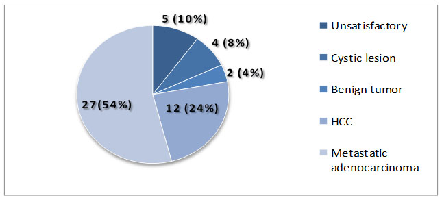
Figure 1. Pie diagram showing distribution of study cases according to FNA cytomorphology
Finally, 8 unsatisfactory, 3 benign and 33 malignant cases were diagnosed in cell block preparations. Among 33 malignant cases 6 were diagnosed as hepatocellular carcinoma (HCC) and 27 were diagnosed as metastatic adenocarcinoma (Figure-2).
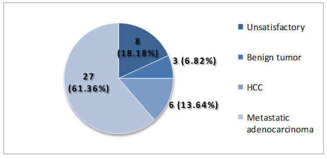
Figure 2. Pie diagram showing distribution of 44 cases according to combined cell block preparations.
36 cases were conclusive on both cytomorphology and cell block preparations. On evaluation of cytomorphological diagnosis of 36 cases, 33 were true positive diagnosis, 2 were true negative diagnosis, 1 was false positive diagnosis and there was no false negative diagnosis (Table II). Sensitivity, specificity, PPV, NPV and accuracy of FNAC in the diagnosis of malignant focal liver lesions were100%, 66.67%, 97.06 %, 100 % & 97.22 %, respectively.
Table II: Statistical evaluation of cytomorphological diagnosis of 36 conclusive cases.
| Combined cell block preparations (H&E and IHC) | Cytomorphological diagnosis | |
| Disease positive (Malignant) | Disease negative(Benign) | |
| Positive(Malignant) 33 | TP 33 | FP 1 |
| Negative(Benign) 3 | FN 0 | TN 2 |
| Total 36 | 33 | 3 |
TP= True positive, TN= True negative, FP= False positive, FN= False negative
33 cases were diagnosed as malignant by both FNAC and cell block preparations. On evaluation of cytomorphological diagnosis, 4 were true positive, 23 were true negative, 4 were false positive and 2 were false negative in the detection of HCC (Table III). Sensitivity, specificity, PPV, NPV and accuracyof FNAC in the detection of HCC were 66.67%, 85.18%, 50%, 92% and 81.82%, respectively.
Table III: Statistical evaluation of cytomorphological diagnosis in the detection of HCC.
| Combined cell block preparations (H&E and IHC) | Cytomorphological diagnosis | |
| Disease positive (HCC) | Disease negative
(Non HCC) |
|
| Positive (HCC) 6 | TP 4 | FP 4 |
| Negative (Non HCC) 27 | FN 2 | TN 23 |
| Total 33 | 6 | 27 |
TP= True positive, TN= True negative, FP= False positive, FN= False negative
Discussion
In the present study, USG guided FNAC was compared with cell block preparations (H&E and immunohistochemistry) in differentiation of focal liver lesions. FNA smears were available in all the 50 cases, but cell blocks were available in 44 cases.
Age of the study patients ranged from 15 to 80 years with a mean of 53.58 years. Nazir et al. (2010) and Kuo et al. (2004) showed 55 and 58.1 years as mean age in their studies which are close to the mean age of present study.7,8 Highest number of patients (26%) was in the age group of 51-60 years in our study. Nazir et al. (2010) reported that maximum number of cases was seen between 55-65 years of age which is nearly similar to present study.7 Out of 50 cases, 33 (66%) were male and 17 (34%) were female with male to female ratio of 1.94:1. Similar findings were reported by Swamy et al. (2011).9 Nazir et al. (2010) showed a male to female ratio of 1.7:1 which is also close to present study.7
Out of 50 cases, 5 (10%) cases were unsatisfactory, 4 (8%) cases were cystic lesion, 2 (4%) cases were benign tumor and 39 (78%) cases were malignant tumor on cytomorphology. Further categorization of benign tumors was not done as in Khurana et al. (2009).6 Among 39 malignant cases, 12 (30.77%) cases were HCC and 27 (69.23%) cases were metastatic carcinoma. All the cases of metastatic carcinoma were adenocarcinomas. Nearly similar findings were found on cytomorphology in the study of Mohmmed et al. (2012), Nazir et al. (2010), Khurana et al. (2009) and Ceyhan et al. (2006).6,7,10,11 Ozkara et al. (2012) found 9.9% of cases as unsatisfactory on cytomorphology which is similar to the unsatisfactory smear (10%) of the present study.12
In final diagnosis of 44 cases by combined cell block preparations (H&E and immunohistochemistry), 8 (18.18%) were unsatisfactory, 3 (6.82%) were benign tumors and 33 (75%) were malignant tumors. Nazir et al. (2010) reported 85% cases as malignant which is nearly close to the malignant cases found in the present study.7 But Mohmmed et al. (2012) showed 39% cases as malignant which is lower and Khurana et al. (2009) showed 93.75% cases as malignant which is higher than that of present study.6,10 Among the malignant lesions, 6 (18.18%) were HCC and 27 (81.82%) were metastatic adenocarcinoma in our study. Khurana et al. (2009) found 17.78% cases as HCC and 82.22% cases as metastatic tumor which are concordant with the present study.6
The sensitivity, specificity, and accuracy of USG guided FNAC in the evaluation of focal liver lesions were 100%, 66.67% and 97.22%, respectively. Sensitivity of the present study (100%) is similar or close to the sensitivity of studies done by Khurana et al. (2009), Nazir et al. (2010), Swamy et al. (2011) and Mohmmed et al. (2012).6,7,9,10 Specificity of the present study (66.67%) has concordance with the specificity found by Mohmmed et al. (2012).10 The specificity shown by Khurana et al. (2009), Nazir et al. (2010) and Swamy et al. (2011) has discordance with that of current study.6,7,9The present study showed an accuracy of 97.22% which is similar to that of Nazir et al. (2010) and Swamy et al. (2011).7,9
The sensitivity, specificity, and accuracy of FNAC in the detection of HCC were 66.67%, 85.18% and 81.82% respectively in our study. Sensitivity of FNAC in the detection of HCC described by Ozkara et al. (2013) was 68.2% which is similar to the sensitivity of present study.12 Khurana et al. (2009) and Nazir et al. (2010) showed the sensitivity in the detection of HCC as 72.3% and 96% respectively which are higher than the sensitivity of present study.6,7 Specificity and accuracy showed by Nazir et al. (2010) were 100% and 97.5% respectively which are also higher than those of the present study.7
Conclusion
FNAC of focal liver lesions has high sensitivity and accuracy in the detection of malignancy but it has low sensitivity in the detection of HCC. No significant complication was observed during aspiration. FNAC is a relatively safe, quick, cost effective and patient compliant procedure which has high accuracy in the differentiation between benign and malignant focal liver lesions. Simultaneous cell block preparations can improve the efficacy of FNAC in the subtyping of malignancy.
References
![]()
Correlation of Ki-67 Proliferating Index with Histological Stage and Grade in Colorectal Carcinoma
*Sultana S,1 Islam N,2 Kabir E,3 Akhter S,4 Paul R,5 Shirin A,6 Khan AA,7 Jahan N8
Abstract
Colorectal carcinoma is the most common cancer of gastrointestinal tract. It is the 3rd most commonly diagnosed cancer and the 3rd leading cause of cancer death. The growth of tumor in colorectal carcinoma is highly variable and its histological grading and staging has important role in diagnosis, treatment and overall prognosis. To observe the Ki-67 expression in colorectal carcinoma and find out the possible correlation of Ki-67 proliferating index with histological grading and Duke’s staging. This cross sectional study was conducted at Sir Salimullah Medical College from July 2014 to June 2016. 98 patients with colorectal carcinoma enrolled in this study by purposive sampling. The H&E staining was done on paraffin embedded tissue sample. Ki-67 expression by IHC method. Ki-67 is a proliferation associated nuclear antigen which can be recognize by MIB-1 monoclonal antibody, correlate with histological staging and grading in colorectal carcinoma. Then tumours were graded according to WHO grading criteria and pathological staging was done according to Duke’s staging system and immunohistochemical staining for Ki-67 antigen expression. Results were subjected to statistical analysis. The results were considered to be significant when the P< 0.05. Ki-67 proliferative index was high in well and moderately differentiated adenocarcinoma but low in poorly differentiated which is statistically significant (p <0.05). Ki-67 expression was high in early Duke’s stage A and B but low expression in advanced Duke’s stage C (p>0.05). The result of this study will enlighten the clinician regarding the need for doing Ki-67 in colorectal carcinoma which would contribute to better understanding of the treatment as well as prognosis.
[Journal of Histopathology and Cytopathology, 2018 Jul; 2 (2):125-133]
Key words: Colorectal carcinoma, Immunohistochemistry, Ki-67
*For correspondence
Introduction
Colorectal carcinoma is the most common malignancy of gastrointesinal (GI) tract and is a major cause of morbidity and mortality worldwide.1 Colorectal cancer accounts for 10% of all cancers and it is the 2nd leading cause of death from malignancy in the industrialized world.2 There are nearly one million new cases of colorectal cancer diagnosed worldwide each year and half a million death.3 In 2013, there were an estimated 1,177,556 people living with colon and rectal cancer in the United States and the number of new cases of colon and rectal cancer was 41.0 per 100,000 men and women per year.4 Regarding age incidence of colorectal carcinoma, recent reports show that in the USA it was the most frequent form of cancer among the person aged between 60-70 years and fewer than 20% of cases occurs before the age 50.5 The incidence of colorectal cancer in Bangladesh is exactly not known but estimated population are approximately 15,10.1%.6 The distribution of colorectal carcinoma worldwide seems to be related to industrialization and socioeconomic standard and the incidence rate is higher in industrialized countries including Western Europe, Scandinavia and North America, whereas in the developing countries (sub-Saharan, Africa and Asia) the incidences appear to be lower.7
There are several staging system for colorectal carcinoma among these TNM and Duke’s staging systems are the most common way of staging and grading of colorectal carcinoma.8 The American Joint Committee (AJC) and the Union for International Cancer control (UICC) joined to produce the TNM system, which attempt to record clinical and pathological data, guide therapy and forecast prognosis, all in one.9 Whereas the classification into Duke’s stage A,B,C cases is the measurement of the boundaries reached and both methods permit the grouping of cases into favorable and unfavorable outcome.10 Histologically the tumor is graded according to WHO grading criteria as well differentiated, moderately differentiated and poorly differentiated and the histological appearance of colorectal carcinoma may vary considerably with its major importance being related to prognosis.10
The use of monoclonal antibodies raised against specific antigens associated with the cell proliferation.11 Ki-67 is a proliferation associated nuclear antigen expressed in all cycling cell except resting cell in the G0 phase and it reflects cell in the S/G2+M phases in particularly.12 MIB-1 is a monoclonal antibody and it recognizes the Ki67 nuclear antigen in the formalin fixed paraffin embedded tissue section.13 Ki-67 expression is estimated as the percentage of tumors cells positively stained by the antibody with nuclear staining.12 The importance of Ki-67 as an indicator of tumor behavior and in colorectal cancer this index may be used as a marker of prognosis.12
The proliferative activity as measured by Ki-67 antibody is closely associated with histological grade and stage.2 In 2008 Uzma Nabi, Nagi A H and Waqas Sami, Department of Pathology, University of Health Sciences, Lahore, Pakistan conducted a study on Ki-67 proliferating index and histological stage and grade of colorectal carcinoma and observed that proliferative index is high in well and moderately differentiated adenocarcinoma and in an early Duke’s stage (A or B).2 But Ki-67 proliferating activity is low in poorly differentiated tumour and in an advanced Duke’s stage C.13,8 But there are some studies of Lanza, Cavazzinil in 1990 and Yokoyama N, Okomoto H in Japan in 2005 contradicting the above mentioned association of Ki-67 versus grading and staging of colorectal carcinoma, they concluded that proliferating index of Ki-67 was increasing with increasing grade, stage.14, 15
Methods
This cross sectional study was conducted among the 98 histopathologically diagnosed patients having colorectal carcinoma over a period of two years in the department of surgery, Sir Salimullah Medical College. Study population were the patients having colorectal cancer underwent surgical treatment in the department of surgery of Sir Salimullah Medical College. The proliferative activity as measured by Ki-67 antibody is closely associated with histological grading and staging of colorectal carcinoma. The representative sections were submitted for Immunohistochemical staining. The Ki-67 immunostaining were performed according to manufacture’s recommendation, using the MIB-1 clone (DAKO, Carpenteria, CA & Ventena Medical System, Tucson, AZ). Ki-67 immunostainined slides were examined via light microsccopy. Positive Ki-67 staining was observed brown granular nuclear staning. For Ki-67 scoring the most positive area of the tumor was selected avoiding foci of inflammation. The number of positive nuclei were counted in 500 tumor cells in a high power field. The average of the counts over the same slides was taken and expressed as the percentage of Ki-67 positive cells in the tumor.
Statistical analysis were performed in SPSS statistical software program, Version 17.0. To correlate histological grading and staging of colorectal carcinoma with Ki-67 proliferating index were performed with Mann- Whitney U test. The result were consider to be significant when P<0.05. One way ANOVA followed by Bonferroni test was performed to compare between groups.
Results
98 cases were included in the present study. Age incidence ranged from 28-78 years and their mean ± SD 47.38 ± 10.37. Maximum patients (30.6 %) were found in 41-50 years age group where M: F was 1.72: 1 (Table I). Regarding site of tumor more than 50% of patient had tumors in the left side of colon and in the rest of the cases tumors were present in caecum (26.5%; Table-II). 98 cases having different sizes of tumors and in most of the cases fifty nine cases (60.2%) tumors size were 3-4 cm and their mean ± SD 4.8 ± 1.8 (Table-III). Different morphological types of tumor were observed in present study. In maximum forty cases (40.8%) the morphological types of tumors were ulcerative and only five cases (5.3%) tumors were infiltrative type. Rests were annular (33.5%) and polypoid (20.3%; Table IV).
In the present study, tumors were graded according to WHO grading system into well differentiated, moderately differentiated, poorly differentiated and grouped into A, B, C accordingly. The maximum cases 69 (70.4%) of colorectal carcinoma were well differentiated and their mean Ki-67 proliferating index was 47.83 ± 15.23. There were significant differences among the groups (A vs B vs C), when mean proliferating index of Ki-67 were compared among the three groups. The result was found statistically significant (P<0.05). However when compared in between groups only A vs C (between well differentiate & poorly differentiate) groups were found also statistically significant (P<0.05). In the present study, it was also observed that with increasing grade, Ki-67 proliferating index decreases (Table V) .
Regarding staging of the tumor (Duke’s staging) where maximum cases (44.8%) were in stage B1 and their mean Ki-67 proliferating index was 46.25±14.06. There was no significant differences among the stages and the result was not statistically significant (P>0.05). In the present study it was also observed that with increasing stage of the tumors, there was decreasing Ki-67 proliferating index (Table VI).
Table I: Distribution of patients according to age group with male female ratio (n=98)
| Age groups | Frequency | M: F | Percentage | ||
| Total | Male | Female | |||
| ≤30
|
10 | 7 | 3 | 2.3:1 | 10.2 |
| 31-40 | 23 | 13 | 10 | 1.3: 1 | 23.5 |
|
41-50 |
30 |
19 |
11 |
1.72: 1 |
30.6 |
|
51-60 |
26 |
17 |
9 |
1.88: 1 |
26.5 |
|
≥60 |
9 |
9 |
0 |
9.00: 0 |
9.2 |
|
Total |
98 |
||||
|
Mean ± SD
|
47.01± 10.99 |
||||
| Range (Min-Max) 28 – 78
|
|||||
Table II: Distribution of tumors according to site (n=98)
| Site of tumor | Frequency (%) |
| Left side of colon | |
| Sigmoid colon | 34 (34.7) |
| Transverse colon | 24 (24.6) |
| Rectum | 14 (14.7) |
| Right side of colon | |
| Caecum | 26 (26.5) |
| Total | 98 (100) |
Table III: Distribution of patients according to tumor size (n=98)
| Tumor size (cm) | Frequency (%) |
| 1 – 2 | 14 (14.3) |
| 3 – 4 | 59 (60.2) |
| 5 – 6 | 19 (19.4) |
| Mean ± SD | 4.8 ± 1.8 |
| Total | 98 (100) |
Table IV: Distribution of patients according to morphological types of tumor (n=98)
| Morphology | Frequency (%) |
| Ulcerative | 40 (40.8) |
| Annular | 33 (33.5) |
| Polypoid | 20 (20.3) |
| Infiltrating | 5 (5.3) |
| Total | 98 (100) |
Table V: Relation of Ki-67 proliferating index with histological grading (n=98)
| Grading | Frequency
n(%) |
Ki-67 expression
(Mean ± SD) |
P |
| Well differentiated (A) | 69 (70.4) | 47.83± 15.23 | |
| Moderate differentiate (B) |
15(15.3) | 46.33 ± 18.07 | |
| Poor differentiated (C) | 14(14.3) | 35.35 ± 11.17 | |
| Statistical analysis | |||
| A vs B vs C | 0.023* | ||
| A vs B | 1.000ns | ||
| A vs C | 0.019* | ||
| B vs C | 0.162 ns |
ANOVA followed by Bonferroni test was performed to compare between groups
Table VI: Relation of Ki-67 proliferating index with histological stage (n=98)
| Duke’s staging
|
Frequency
n (%) |
Ki-67 expression
(Mean ± SD) |
p |
| Stage A | 3 (3.3%) | 53.33 ± 20.81 | 0.727ns |
| Stage B1 | 44 (44.8) | 46.25 ± 14.06 | |
| Stage B2 | 20(20.5) | 43.25 ± 14.06 | |
| Stage C1 | 14(14.8) | 44.64 ± 21.07 | |
| Stage C2 | 17(17.5) | 42.05 ± 13.69 |
ANOVA test was done to measure the level of significance.

Figure 1. Photomicrograph of histopathological section of well differentiated adenocarcinoma of colon (H&E method x100)

Figure 2. Photomicrograph of well differentiated adenocarcinoma stained with Ki-67 immunostain showing high proliferative Index(x100).

Figure 3. Photomicrograph of histopathological section of poorly differentiated adenocarcinoma of colon ( H& E method x400).

Figure 4. Photomicrograph of poorly differentiated adenocarcinoma stained with Ki-67 immunostain showing low Proliferative Index (x400).
Discussion
Colorectal cancer (CRC) is one of the most common malignancies and a leading cause of cancer death worldwide.16 The incidence of cancer colon and rectum was 41.0 per 100,000 men and women per year and the number of deaths was 15.1 per 100,000 men and women per year in Bangladesh.17 Management of colorectal carcinoma depends on a number of morphological and biological factors which include the pathological tumor stage (including involvement of lymph nodes, breach of serosa, distant spread etc.), primary tumor characteristics (including depth of tumor penetration in the bowel wall, histological subtype, histological grade and differentiation, venous and lymphatic invasion, perineural invasion and lymphocytic infiltration), status of surgical resection margins (free or involved).18 With assessment of tumor cell proliferation may predict tumor behavior.19 The aim of this study was to evaluate the proliferating index (PI) in formalin fixed, paraffin embedded tissue section of colorectal carcinoma, using monoclonal MIB-1 antibody (Ki-67) and to assess the relationship between proliferative index (PI) and various pathological findings in colorectal carcinoma including histological grade, and stage.
In the present study, the mean age of the patients was 47.01± 10.99 years and the highest number of malignant cases were seen in the 4th and 5th decades. Male female ratio was 1.72:1 and 1.88:1 in 4th and 5th decades accordingly. So, in present study, male were predominant than female. These findings were similar to other studies.20, 21
In 34.7% patients the tumors were located in sigmoid colon followed by caecum (26.5%), transverse colon (24.5%) and rectum (14.3%). As per gross morphological type of cancer, maximum 40.8% were ulcerative and 33.5% were annular type and rest were polypoid (20.3%) and infiltrating type (5.3%). In one study, colon was more commonly affected site compared to rectum and most of the lesions were ulcerative.22 In another study rectum was the most common affected site and predominant lesions were annular.23
In present study, 60.2% of malignant cases had tumors size between 3-4 cm and 19.4% malignant cases had tumors size 5-6 cm. In a study in 2011 by Kornprat showed that maximum size of the tumor in colon cancer were in between 4.5 to 6.5 cm in majority of cases.24 In 2013 ASCO annual meeting showed a report where a study conducted on tumor size in colorectal cancer found that in majority of the cases the tumors sizes were in between 4-6 cm in colorectal carcinoma.25
In this present study it was observed that the Ki-67 labelling index was high in grade I and grade II compared to grade III. These results showed that proliferating index was low in poorly differentiated tumor compared to well and moderately differentiated tumor. When Ki-67 proliferating index was compared among three groups a statistically significant correlation was found in present study (p<0.05). These findings are similar to studies in Japan2 Pakistan12 and Finland.26 On the other hand, some of the studies showed contradictory result that Ki-67 proliferating index increased with increasing histological grade.26,27
In the present study, it was observed that Ki 67 labeling index was high in Duke’s stage A and B and tumor in advanced stage (Duke’s C) have a low proliferating index compared to tumors in an early invasive stage (Duke’s A and B). Regarding the correlation of Ki-67 LI with staging, no statistically significant correlation was found. This result was similar with other studies in which they concluded that Ki-67 proliferating index was significantly lower in carcinoma in subserosa or deeper invasion compared to carcinoma with submucosa or muscularis mucosa invasion.2,8,26 In another study contradictory results have been reported on associations of Ki-67 with prognosis and survival of colorectal tumors with a low proliferation index in Duke’s A and B tumors to be associated with survival impairment compared to those with high values.28 Tumors with high proliferative activity are known to be most responsive to radiotherapy and Willett and collaborators showed that radiation eradicates preferentially rapidly dividing cells in rectal cancer, whereas populations with slow proliferation show greater radioresistance.29
Conclusion
It is concluded that Ki-67 labeling index is high in well to moderately differentiated adenocarcinomas in an early Duke’s stage A or B compared to poorly differentiated adenocarcinomas, and in an advanced Duke’s stage C. Thus Ki-67 proliferating index can be useful in a patient with colorectal carcinoma as a ancillary diagnostic support. Moreover, it may help in the prognostic evaluation of patient, survival as well as in considering them for post surgical treatment.
References